The CD11b/ITGAM Antibody (CAB1581) is a high-quality antibody developed for reliable detection and analysis of target proteins. This antibody, produced using rabbit immunization, exhibits high specificity and reactivity towards human samples, making it a reliable choice for Western blot applications.Integrin alpha M functions as a receptor for various ligands, mediating cell-cell and cell-extracellular matrix interactions. Its role in immune cell migration and phagocytosis highlights its importance in inflammatory responses and host defense mechanisms.
This antibody is validated for use in WB, IHC-P, IF/ICC, ELISA applications and has demonstrated reactivity against Human, Mouse, Rat samples.
Product Name:
CD11b/ITGAM Antibody
SKU:
CAB1581
Size:
20μL, 100μL
Reactivity:
Human, Mouse, Rat
Conjugate:
Unconjugated
Immunogen:
Recombinant protein (or fragment).This information is considered to be commercially sensitive.
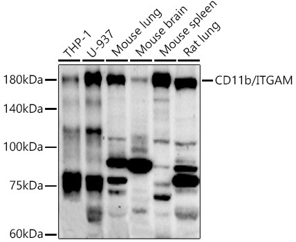
THP-1, U-937, Mouse lung, Mouse brain, Mouse spleen, Rat lung
Cellular Localization:
Membrane, Single-Pass Type I Membrane Protein.
Calculated MW:
127kDa
Observed MW:
170kDa
This gene encodes the integrin alpha M chain. Integrins are heterodimeric integral membrane proteins composed of an alpha chain and a beta chain. This I-domain containing alpha integrin combines with the beta 2 chain (ITGB2) to form a leukocyte-specific integrin referred to as macrophage receptor 1 ('Mac-1'), or inactivated-C3b (iC3b) receptor 3 ('CR3'). The alpha M beta 2 integrin is important in the adherence of neutrophils and monocytes to stimulated endothelium, and also in the phagocytosis of complement coated particles. Multiple transcript variants encoding different isoforms have been found for this gene.
Purification Method
Affinity purification
Gene ID
3684
RRID
AB_2763232
Buffer Information
Store at -20℃. Avoid freeze / thaw cycles. Buffer: PBS with 0.09% Sodium azide,50% glycerol,pH7.3.
Western blot analysis of various lysates using CD11b/ITGAM Rabbit pAb (CAB1581) at 1:500 dilution. Secondary antibody: HRP-conjugated Goat anti-Rabbit IgG (H+L) (CABS014) at 1:10000 dilution. Lysates/proteins: 25μg per lane. Blocking buffer: 3% nonfat dry milk in TBST. Detection: ECL Enhanced Kit (AbGn00021). Exposure time: 90s.
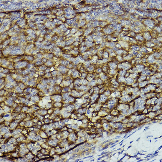
Immunohistochemistry analysis of paraffin-embedded Human tonsil using CD11b/ITGAM Rabbit pAb (CAB1581) at dilution of 1:100 (40x lens). High pressure antigen retrieval performed with 0.01M Citrate buffer (pH 6.0) prior to IHC staining.
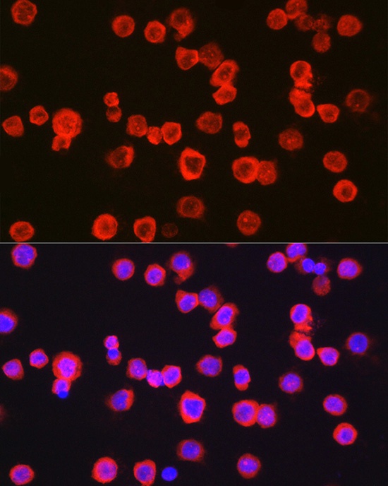
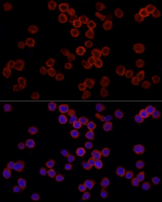
Immunofluorescence analysis of TF-1 cells using CD11b/ITGAM Rabbit pAb (CAB1581) at dilution of 1:100 (40x lens). Secondary antibody: Cy3-conjugated Goat anti-Rabbit IgG (H+L) (CABS007) at 1:500 dilution. Blue: DAPI for nuclear staining.
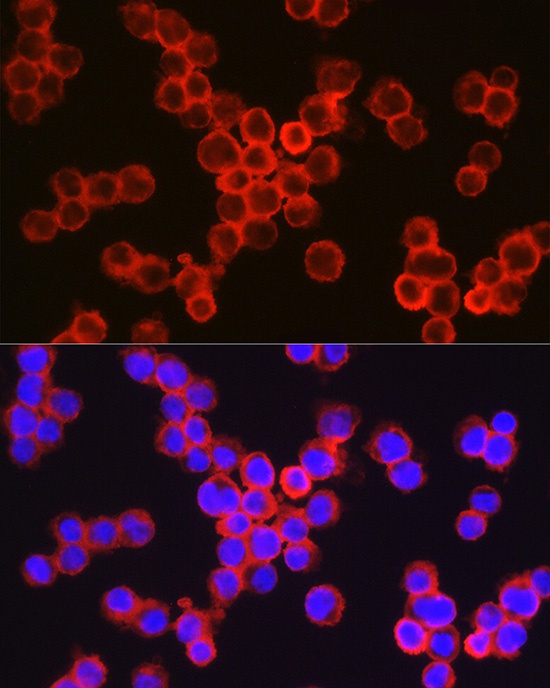
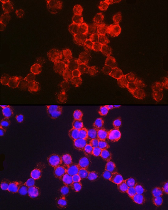
Immunofluorescence analysis of THP-1 cells using CD11b/ITGAM Rabbit pAb (CAB1581) at dilution of 1:100 (40x lens). Secondary antibody: Cy3-conjugated Goat anti-Rabbit IgG (H+L) (CABS007) at 1:500 dilution. Blue: DAPI for nuclear staining.
Immunofluorescence analysis of THP-1 cells using CD11b/ITGAM Rabbit pAb (CAB1581) at dilution of 1:100 (40x lens). Secondary antibody: Cy3-conjugated Goat anti-Rabbit IgG (H+L) (CABS007) at 1:500 dilution. Blue: DAPI for nuclear staining.
Immunofluorescence analysis of TF-1 cells using CD11b/ITGAM Rabbit pAb (CAB1581) at dilution of 1:100 (40x lens). Secondary antibody: Cy3-conjugated Goat anti-Rabbit IgG (H+L) (CABS007) at 1:500 dilution. Blue: DAPI for nuclear staining.


")
")
