The IRAK2 Monoclonal Antibody (CAB4655) is a high-quality antibody developed for reliable detection and analysis of target proteins. This antibody, produced in rabbits, is highly specific for IRAK2 and has been validated for use in various applications such as Western blotting and immunohistochemistry.IRAK2, also known as interleukin-1 receptor-associated kinase 2, is a crucial mediator of Toll-like receptor and interleukin-1 receptor signaling pathways, leading to the activation of NF-κB and inflammatory responses. Dysregulation of IRAK2 has been implicated in various inflammatory diseases, making it a promising target for therapeutic intervention.
This antibody is validated for use in WB, IHC-P, ELISA applications and has demonstrated reactivity against Human, Rat samples.
Product Name:
IRAK2 Monoclonal Antibody
SKU:
CAB4655
Size:
20μL, 100μL
Reactivity:
Human, Rat
Clone Number:
ARC1078
Conjugate:
Unconjugated
Immunogen:
Recombinant protein (or fragment).This information is considered to be commercially sensitive.
Recommended starting concentration is 1 μg/mL. Please optimize the concentration based on your specific assay requirements.
Synonyms:
IRAK-2, IRAK2
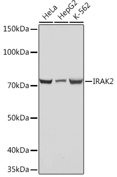
Positive Sample:
HeLa, HepG2, K-562
Cellular Localization:
Cytoplasm, Cytosol, Nucleus, Plasma Membrane.
Calculated MW:
69kDa
Observed MW:
69kDa
IRAK2 encodes the interleukin-1 receptor-associated kinase 2, one of two putative serine/threonine kinases that become associated with the interleukin-1 receptor (IL1R) upon stimulation. IRAK2 is reported to participate in the IL1-induced upregulation of NF-kappaB.
Purification Method
Affinity purification
Gene ID
3656
RRID
AB_2863318
Buffer Information
Store at -20℃. Avoid freeze / thaw cycles. Buffer: PBS containing 50% glycerol and 0.05% BSA, preserved with proclin300 or sodium azide, pH 7.3.
Western blot analysis of various lysates using IRAK2 Rabbit mAb (CAB4655) at 1:1000 dilution. Secondary antibody: HRP-conjugated Goat anti-Rabbit IgG (H+L) (CABS014) at 1:10000 dilution. Lysates/proteins: 25μg per lane. Blocking buffer: 3% nonfat dry milk in TBST. Detection: ECL Basic Kit (AbGn00020). Exposure time: 1s.
Immunohistochemistry analysis of paraffin-embedded Rat kidney using IRAK2 Rabbit mAb (CAB4655) at dilution of 1:100 (40x lens). Microwave antigen retrieval performed with 0.01M PBS Buffer (pH 7.2) prior to IHC staining.