The IRF6 Monoclonal Antibody (CAB3209) is a high-quality antibody developed for reliable detection and analysis of target proteins. This antibody, generated in rabbits, is highly specific to human IRF6 samples and has been validated for use in Western blot applications.IRF6 is essential for proper cell differentiation and proliferation, making it a crucial regulator of tissue development and repair. Dysregulation of IRF6 has been linked to a variety of developmental disorders, including orofacial clefts and skin abnormalities.
This antibody is validated for use in WB, IF/ICC, IP, ELISA applications and has demonstrated reactivity against Human, Mouse, Rat samples.
Product Name:
IRF6 Monoclonal Antibody
SKU:
CAB3209
Size:
20μL, 100μL
Reactivity:
Human, Mouse, Rat
Clone Number:
ARC1928
Conjugate:
Unconjugated
Immunogen:
Synthetic peptide. This information is considered to be commercially sensitive.
0.5μg-4μg antibody for 200μg-400μg extracts of whole cells
ELISA
Recommended starting concentration is 1 μg/mL. Please optimize the concentration based on your specific assay requirements.
Synonyms:
LPS, PIT, PPS, VWS, OFC6, PPS1, VWS1, IRF6
Positive Sample:
293T, HepG2, A-549, Mouse lung, Mouse kidney, Rat lung, Rat liver
Cellular Localization:
Cytoplasm, Nucleus.
Calculated MW:
53kDa
Observed MW:
68kDa
This gene encodes a member of the interferon regulatory transcription factor (IRF) family. Family members share a highly-conserved N-terminal helix-turn-helix DNA-binding domain and a less conserved C-terminal protein-binding domain. The encoded protein may be a transcriptional activator. Mutations in this gene can cause van der Woude syndrome and popliteal pterygium syndrome. Mutations in this gene are also associated with non-syndromic orofacial cleft type 6. Alternate splicing results in multiple transcript variants.
Purification Method
Affinity purification
Gene ID
3664
RRID
AB_2863026
Buffer Information
Store at -20℃. Avoid freeze / thaw cycles. Buffer: PBS containing 50% glycerol and 0.05% BSA, preserved with proclin300 or sodium azide, pH 7.3.
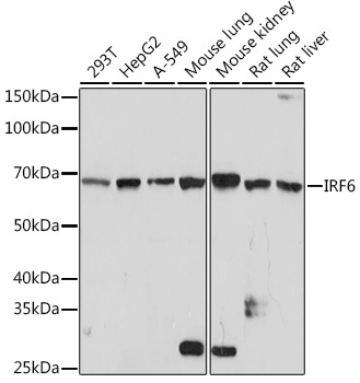
Western blot analysis of various lysates using IRF6 Rabbit mAb (CAB3209) at 1:1000 dilution. Secondary antibody: HRP-conjugated Goat anti-Rabbit IgG (H+L) (CABS014) at 1:10000 dilution. Lysates/proteins: 25μg per lane. Blocking buffer: 3% nonfat dry milk in TBST. Detection: ECL Basic Kit (AbGn00020). Exposure time: 3min.
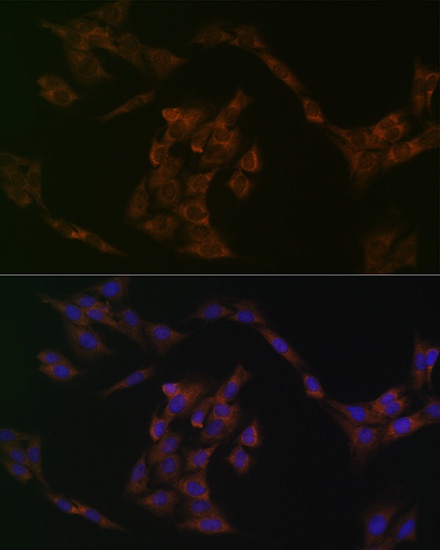
Immunofluorescence analysis of C6 cells using IRF6 Rabbit mAb (CAB3209) at dilution of 1:100 (40x lens). Secondary antibody: Cy3-conjugated Goat anti-Rabbit IgG (H+L) (CABS007) at 1:500 dilution. Blue: DAPI for nuclear staining.
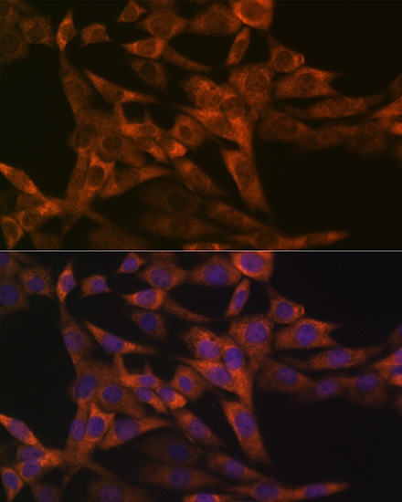
Immunofluorescence analysis of NIH-3T3 cells using IRF6 Rabbit mAb (CAB3209) at dilution of 1:100 (40x lens). Secondary antibody: Cy3-conjugated Goat anti-Rabbit IgG (H+L) (CABS007) at 1:500 dilution. Blue: DAPI for nuclear staining.
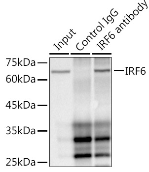
Immunoprecipitation analysis of 300 μg extracts of HepG2 cells using 3 μg IRF6 antibody (CAB3209). Western blot was performed from the immunoprecipitate using IRF6 antibody (CAB3209) at a dilution of 1:1000.