The JAB1/CSN5/COPS5 Monoclonal Antibody (CAB4087) is a high-quality antibody developed for reliable detection and analysis of target proteins. The protein encoded by this gene is one of the eight subunits of COP9 signalosome, a highly conserved protein complex that functions as an important regulator in multiple signaling pathways. The structure and function of COP9 signalosome is similar to that of the 19S regulatory particle of 26S proteasome. COP9 signalosome has been shown to interact with SCF-type E3 ubiquitin ligases and act as a positive regulator of E3 ubiquitin ligases. This protein is reported to be involved in the degradation of cyclin-dependent kinase inhibitor CDKN1B/p27Kip1. It is also known to be an coactivator that increases the specificity of JUN/AP1 transcription factors.
This antibody is validated for use in WB, IHC-P, IF/ICC, ChIP, ELISA applications and has demonstrated reactivity against Human, Mouse, Rat samples.
Product Name:
JAB1/CSN5/COPS5 Monoclonal Antibody
SKU:
CAB4087
Size:
100μL, 20μL
Reactivity:
Human, Mouse, Rat
Clone Number:
ARC0895
Conjugate:
Unconjugated
Immunogen:
Recombinant protein (or fragment).This information is considered to be commercially sensitive.
Tested Applications:
WBIHC-PIF/ICCChIPELISA
Recommended Dilution:
WB
1:500 - 1:2000
IHC-P
1:50 - 1:200
IF/ICC
1:50 - 1:200
ELISA
Recommended starting concentration is 1 μg/mL. Please optimize the concentration based on your specific assay requirements.
ChIP
5μg antibody for 10μg-15μg of Chromatin
Synonyms:
CSN5, JAB1, SGN5, MOV-34, JAB1/CSN5/COPS5
Positive Sample:
HeLa, 293T, Mouse testis, Mouse kidney, Rat testis, Rat brain
The protein encoded by this gene is one of the eight subunits of COP9 signalosome, a highly conserved protein complex that functions as an important regulator in multiple signaling pathways. The structure and function of COP9 signalosome is similar to that of the 19S regulatory particle of 26S proteasome. COP9 signalosome has been shown to interact with SCF-type E3 ubiquitin ligases and act as a positive regulator of E3 ubiquitin ligases. This protein is reported to be involved in the degradation of cyclin-dependent kinase inhibitor CDKN1B/p27Kip1. It is also known to be an coactivator that increases the specificity of JUN/AP1 transcription factors.
Purification Method
Affinity purification
Gene ID
10987
RRID
AB_2863184
Buffer Information
Store at -20℃. Avoid freeze / thaw cycles. Buffer: PBS containing 50% glycerol and 0.05% BSA, preserved with proclin300 or sodium azide, pH 7.3.
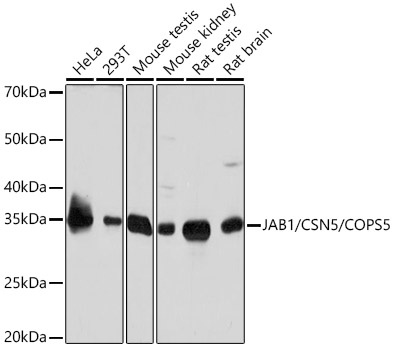
Western blot analysis of various lysates using JAB1/CSN5/COPS5 Rabbit mAb (CAB4087) at 1:1000 dilution. Secondary antibody: HRP-conjugated Goat anti-Rabbit IgG (H+L) (AS014) at 1:10000 dilution. Lysates/proteins: 25μg per lane. Blocking buffer: 3% nonfat dry milk in TBST. Detection: ECL Basic Kit (AbGn00020). Exposure time: 10s.
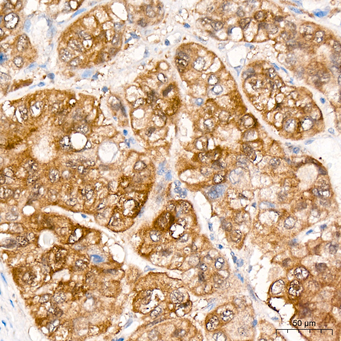
Immunohistochemistry analysis of paraffin-embedded Human liver cancer tissue using JAB1/CSN5/COPS5 Rabbit mAb (CAB4087) at a dilution of 1:100 (40x lens). High pressure antigen retrieval was performed with 0.01 M citrate buffer (pH 6.0) prior to IHC staining.
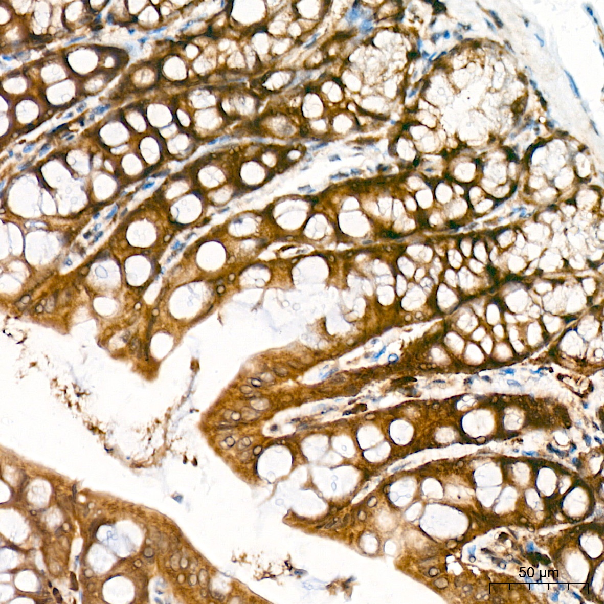
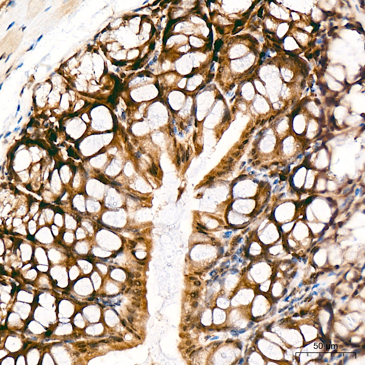
Immunohistochemistry analysis of paraffin-embedded Rat colon tissue using JAB1/CSN5/COPS5 Rabbit mAb (CAB4087) at a dilution of 1:100 (40x lens). High pressure antigen retrieval was performed with 0.01 M citrate buffer (pH 6.0) prior to IHC staining.
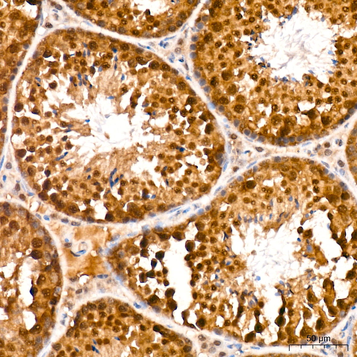
Immunohistochemistry analysis of paraffin-embedded Mouse testis tissue using JAB1/CSN5/COPS5 Rabbit mAb (CAB4087) at a dilution of 1:100 (40x lens). High pressure antigen retrieval was performed with 0.01 M citrate buffer (pH 6.0) prior to IHC staining.
Immunohistochemistry analysis of paraffin-embedded Mouse colon tissue using JAB1/CSN5/COPS5 Rabbit mAb (CAB4087) at a dilution of 1:100 (40x lens). High pressure antigen retrieval was performed with 0.01 M citrate buffer (pH 6.0) prior to IHC staining.
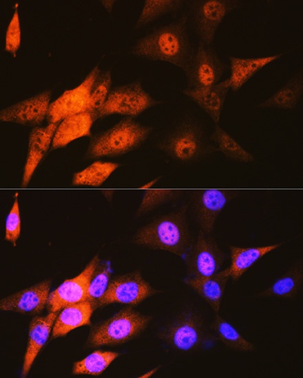
Immunofluorescence analysis of NIH-3T3 cells using JAB1/CSN5/COPS5 Rabbit mAb (CAB4087) at dilution of 1:100 (40x lens). Secondary antibody: Cy3-conjugated Goat anti-Rabbit IgG (H+L) (AS007) at 1:500 dilution. Blue: DAPI for nuclear staining.
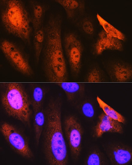
Immunofluorescence analysis of U-2 OS cells using JAB1/CSN5/COPS5 Rabbit mAb (CAB4087) at dilution of 1:100 (40x lens). Secondary antibody: Cy3-conjugated Goat anti-Rabbit IgG (H+L) (AS007) at 1:500 dilution. Blue: DAPI for nuclear staining.