The KDM3A Antibody (CAB11960) is a high-quality antibody developed for reliable detection and analysis of target proteins. This antibody, produced in rabbits, exhibits high specificity for human samples and has been verified for use in Western blot experiments. By binding to the KDM3A protein, this antibody enables precise detection and analysis in various cell types, making it an essential resource for studies in genetics, cancer research, and developmental biology.KDM3A, also known as lysine-specific demethylase 3A, is a histone-modifying enzyme that plays a crucial role in gene expression regulation. Its involvement in chromatin remodeling and transcriptional activation makes it a target of interest in understanding cellular differentiation, proliferation, and disease progression.
This antibody is validated for use in WB, IHC-P, IF/ICC, ELISA applications and has demonstrated reactivity against Human samples.
Product Name:
KDM3A Antibody
SKU:
CAB11960
Size:
20μL, 100μL
Reactivity:
Human
Conjugate:
Unconjugated
Immunogen:
Synthetic peptide. This information is considered to be commercially sensitive.
Recommended starting concentration is 1 μg/mL. Please optimize the concentration based on your specific assay requirements.
Synonyms:
TSGA, JMJD1, JHDM2A, JHMD2A, JMJD1A, KDM3A
Positive Sample:
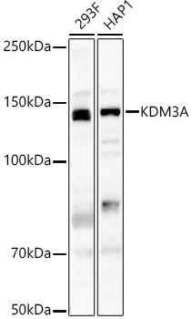
293F, HAP1
Cellular Localization:
Cytoplasm, Nucleus.
Calculated MW:
147kDa
Observed MW:
147kDa
Enables androgen receptor binding activity; histone H3-methyl-lysine-9 demethylase activity; and iron ion binding activity. Involved in several processes, including androgen receptor signaling pathway; formaldehyde biosynthetic process; and histone H3-K9 demethylation. Located in nucleoplasm. Implicated in cervical cancer and colon cancer. Biomarker of Ewing sarcoma; hepatocellular carcinoma; nasopharynx carcinoma; and prostate cancer.
Purification Method
Affinity purification
Gene ID
55818
RRID
AB_2758898
Buffer Information
Store at -20℃. Avoid freeze / thaw cycles. Buffer: PBS containing 50% glycerol, preserved with proclin300 or sodium azide, pH 7.3.
Western blot analysis of various lysates, using KDM3A Rabbit pAb (CAB11960) at 1:500 dilution. Secondary antibody: HRP-conjugated Goat anti-Rabbit IgG (H+L) (CABS014) at 1:10000 dilution. Lysates/proteins: 25μg per lane. Blocking buffer: 3% nonfat dry milk in TBST. Detection: ECL Basic Kit (AbGn00020). Exposure time: 90s.
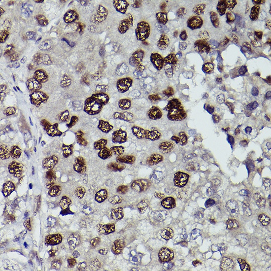
Immunohistochemistry analysis of paraffin-embedded Human liver cancer using KDM3A Rabbit pAb (CAB11960) at dilution of 1:100 (40x lens). High pressure antigen retrieval performed with 0.01M Citrate buffer (pH 6.0) prior to IHC staining.