The KDM4B/JMJD2B Monoclonal Antibody (CAB6670) is a high-quality antibody developed for reliable detection and analysis of target proteins. This highly specific antibody, generated in rabbits, is suitable for use in various applications including Western blot, immunohistochemistry, and immunofluorescence.KDM4B and JMJD2B are members of the Jumonji C domain-containing histone demethylase family, involved in the removal of methyl groups from lysine residues on histone proteins. These proteins play a crucial role in epigenetic regulation, influencing gene expression patterns and cellular differentiation.
This antibody is validated for use in WB, IF/ICC, IP, ChIP, ELISA applications and has demonstrated reactivity against Human, Mouse, Rat samples.
Product Name:
KDM4B/JMJD2B Monoclonal Antibody
SKU:
CAB6670
Size:
20μL, 100μL
Reactivity:
Human, Mouse, Rat
Clone Number:
ARC1416
Conjugate:
Unconjugated
Immunogen:
Synthetic peptide. This information is considered to be commercially sensitive.
0.5μg-4μg antibody for 200μg-400μg extracts of whole cells
ELISA
Recommended starting concentration is 1 μg/mL. Please optimize the concentration based on your specific assay requirements.
ChIP
5μg antibody for 10μg-15μg of Chromatin
Synonyms:
MRD65, JMJD2B, TDRD14B, KDM4B/JMJD2B
Positive Sample:
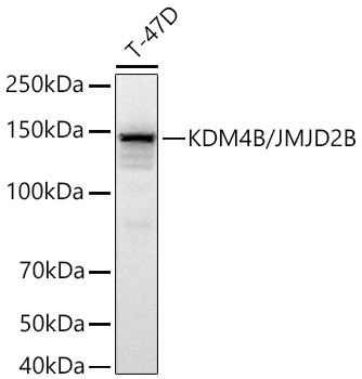
T-47D, HCT 116
Cellular Localization:
Nucleus.
Calculated MW:
122kDa
Observed MW:
150kDa
Enables histone H3-methyl-lysine-36 demethylase activity and histone H3-methyl-lysine-9 demethylase activity. Involved in histone H3-K36 demethylation and histone H3-K9 demethylation. Located in cytosol and nucleoplasm. Implicated in autosomal dominant non-syndromic intellectual disability; breast cancer; colorectal cancer; malignant peripheral nerve sheath tumor; and stomach cancer. Biomarker of several diseases, including alopecia areata; lung cancer; medulloblastoma; prostate cancer; and stomach cancer.
Purification Method
Affinity purification
Gene ID
23030
RRID
AB_2863534
Buffer Information
Store at -20℃. Avoid freeze / thaw cycles. Buffer: PBS containing 50% glycerol and 0.05% BSA, preserved with proclin300 or sodium azide, pH 7.3.
Western blot analysis of lysates from T-47D cells using KDM4B/JMJD2B Rabbit mAb (CAB6670) at 1:7000 dilution incubated at room temperature for 1.5 hours. Secondary antibody: HRP-conjugated Goat anti-Rabbit IgG (H+L) (CABS014) at 1:10000 dilution. Lysates/proteins: 25 μg per lane. Blocking buffer: 3% nonfat dry milk in TBST. Detection: ECL Basic Kit (AbGn00020). Exposure time: 5 s.
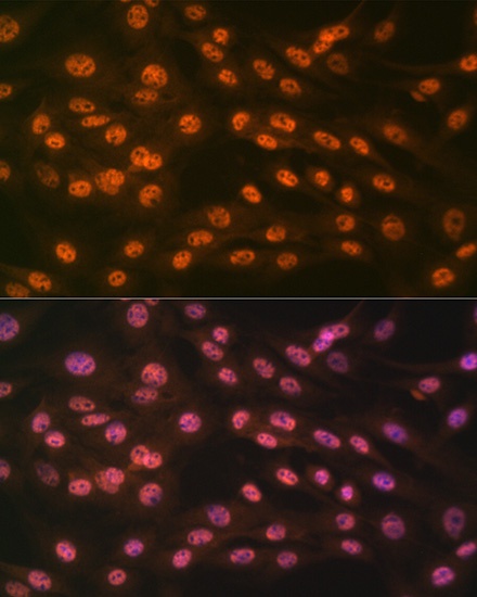
Immunofluorescence analysis of C6 cells using KDM4B/JMJD2B Rabbit mAb (CAB6670) at dilution of 1:100 (40x lens). Secondary antibody: Cy3-conjugated Goat anti-Rabbit IgG (H+L) (CABS007) at 1:500 dilution. Blue: DAPI for nuclear staining.
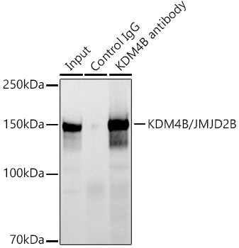
Immunoprecipitation analysis of 300 μg extracts of HCT 116 cells using 3 μg KDM4B/JMJD2B Rabbit mAb (CAB6670). Western blot was performed from the immunoprecipitate using KDM4B/JMJD2B Rabbit mAb (CAB6670) at a dilution of 1:1000.
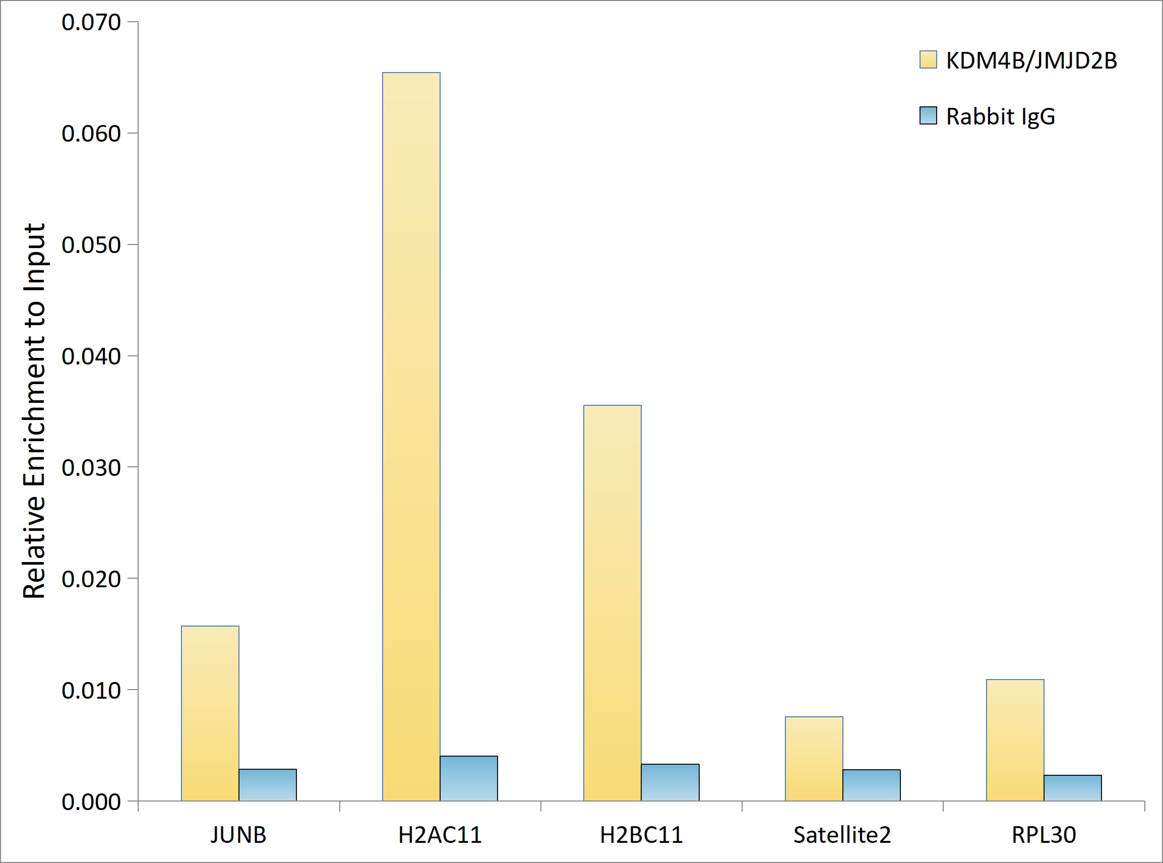
Chromatin immunoprecipitation analysis of extracts of 5637 cells, using KDM4B/JMJD2B Rabbit mAb (CAB6670) and rabbit IgG.The amount of immunoprecipitated DNA was checked by quantitative PCR. Histogram was constructed by the ratios of the immunoprecipitated DNA to the input.