The KIF17 Antibody (CAB16562) is a high-quality antibody developed for reliable detection and analysis of target proteins. Raised in rabbits, this antibody is highly specific for human samples and has been validated for use in Western blot applications. By targeting the KIF17 protein, this antibody allows for the detection and analysis of KIF17 expression in various cell types, making it ideal for investigations in cellular biology and neurology research.
This antibody is validated for use in WB, IHC-P, ELISA, IF-P applications and has demonstrated reactivity against Human, Mouse, Rat samples.
Product Name:
KIF17 Antibody
SKU:
CAB16562
Size:
20μL, 100μL
Reactivity:
Human, Mouse, Rat
Conjugate:
Unconjugated
Immunogen:
Recombinant protein (or fragment).This information is considered to be commercially sensitive.
Recommended starting concentration is 1 μg/mL. Please optimize the concentration based on your specific assay requirements.
Synonyms:
KIF3X, KLP-2, OSM-3, KIF17B, KIF17
Positive Sample:
HeLa, U-87MG, PC-3, Mouse brain, Mouse testis
Cellular Localization:
Cytoplasm, Cytoskeleton.
Calculated MW:
115kDa
Observed MW:
170kDa
Predicted to enable microtubule binding activity and plus-end-directed microtubule motor activity. Predicted to be involved in anterograde dendritic transport of neurotransmitter receptor complex and cell projection organization. Predicted to act upstream of or within microtubule-based process; protein-containing complex localization; and vesicle-mediated transport. Predicted to be located in microtubule cytoskeleton. Predicted to be part of intraciliary transport particle B and kinesin complex. Predicted to be active in cilium; microtubule cytoskeleton; and neuron projection.
Purification Method
Affinity purification
Gene ID
57576
RRID
AB_2770088
Buffer Information
Store at -20℃. Avoid freeze / thaw cycles. Buffer: PBS with 0.01% thimerosal,50% glycerol,pH7.3.
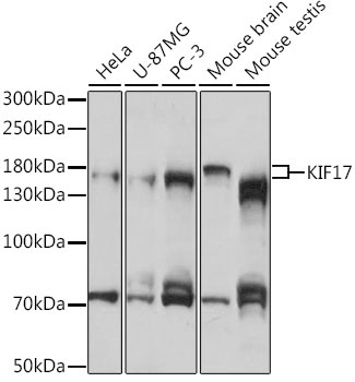
Western blot analysis of various lysates using KIF17 Rabbit pAb (CAB16562) at 1:1000 dilution. Secondary antibody: HRP-conjugated Goat anti-Rabbit IgG (H+L) (CABS014) at 1:10000 dilution. Lysates/proteins: 25μg per lane. Blocking buffer: 3% nonfat dry milk in TBST. Detection: ECL Basic Kit (AbGn00020). Exposure time: 30s.
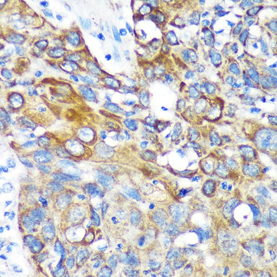
Immunohistochemistry analysis of paraffin-embedded Human lung cancer using KIF17 Rabbit pAb (CAB16562) at dilution of 1:100 (40x lens). Microwave antigen retrieval performed with 0.01M PBS Buffer (pH 7.2) prior to IHC staining.
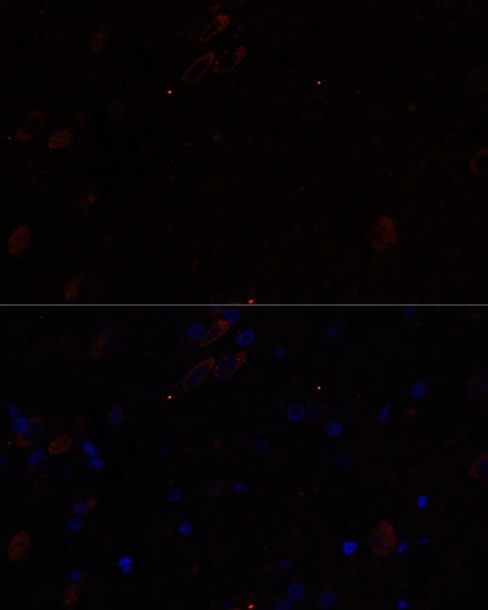
Immunofluorescence analysis of paraffin-embedded rat brain using KIF17 Rabbit pAb (CAB16562) at dilution of 1:100. Secondary antibody: Cy3-conjugated Goat anti-Rabbit IgG (H+L) (CABS007) at 1:500 dilution. Blue: DAPI for nuclear staining.