The KIT Antibody (CAB0357) is a high-quality antibody developed for reliable detection and analysis of target proteins. This antibody, raised in rabbits, targets the CD300A protein, a cell surface molecule that inhibits immune responses. Validated for use in Western blot applications, the CD300A Polyclonal Antibody is highly reactive with human samples, enabling detection and analysis in various cell types.CD300A, also known as an immune inhibitory receptor, plays a critical role in immune homeostasis by regulating inflammation and inhibiting allergic reactions.
This antibody is validated for use in WB, IHC-P, IF/ICC, ELISA applications and has demonstrated reactivity against Human, Mouse, Rat samples.
Product Name:
KIT Antibody
SKU:
CAB0357
Size:
20μL, 100μL
Reactivity:
Human, Mouse, Rat
Conjugate:
Unconjugated
Immunogen:
Recombinant protein (or fragment).This information is considered to be commercially sensitive.
Recommended starting concentration is 1 μg/mL. Please optimize the concentration based on your specific assay requirements.
Synonyms:
PBT, SCFR, C-Kit, CD117, MASTC, CD117/c-Kit
Positive Sample:
Mouse lung
Cellular Localization:
Cell Membrane, Cytoplasm, Single-Pass Type I Membrane Protein.
Calculated MW:
110kDa
Observed MW:
145kDa
This gene encodes a receptor tyrosine kinase. This gene was initially identified as a homolog of the feline sarcoma viral oncogene v-kit and is often referred to as proto-oncogene c-Kit. The canonical form of this glycosylated transmembrane protein has an N-terminal extracellular region with five immunoglobulin-like domains, a transmembrane region, and an intracellular tyrosine kinase domain at the C-terminus. Upon activation by its cytokine ligand, stem cell factor (SCF), this protein phosphorylates multiple intracellular proteins that play a role in in the proliferation, differentiation, migration and apoptosis of many cell types and thereby plays an important role in hematopoiesis, stem cell maintenance, gametogenesis, melanogenesis, and in mast cell development, migration and function. This protein can be a membrane-bound or soluble protein. Mutations in this gene are associated with gastrointestinal stromal tumors, mast cell disease, acute myelogenous leukemia, and piebaldism. Multiple transcript variants encoding different isoforms have been found for this gene.
Purification Method
Affinity purification
Gene ID
3815
RRID
AB_2757143
Buffer Information
Store at -20℃. Avoid freeze / thaw cycles. Buffer: PBS containing 50% glycerol, preserved with proclin300 or sodium azide, pH 7.3.
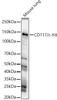
Western blot analysis of lysates from Mouse lung, using CD117/c-Kit Rabbit pAb (CAB0357) at 1:1000 dilution. Secondary antibody: HRP-conjugated Goat anti-Rabbit IgG (H+L) (CABS014) at 1:10000 dilution. Lysates/proteins: 25μg per lane. Blocking buffer: 3% nonfat dry milk in TBST. Detection: ECL Basic Kit (AbGn00020). Exposure time: 20s.
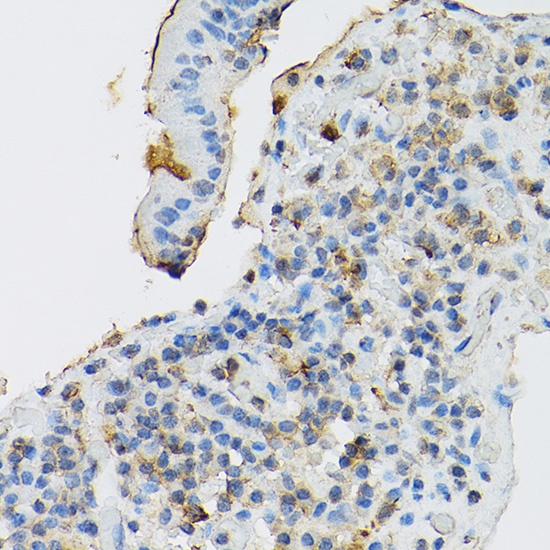
Immunohistochemistry analysis of paraffin-embedded Human gastric cancer using CD117/c-Kit Rabbit pAb (CAB0357) at dilution of 1:100 (40x lens). High pressure antigen retrieval performed with 0.01M Citrate buffer (pH 6.0) prior to IHC staining.
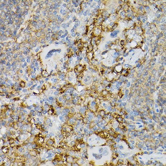
Immunohistochemistry analysis of paraffin-embedded Mouse spleen using CD117/c-Kit Rabbit pAb (CAB0357) at dilution of 1:100 (40x lens). High pressure antigen retrieval performed with 0.01M Citrate buffer (pH 6.0) prior to IHC staining.
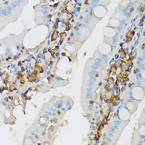
Immunohistochemistry analysis of paraffin-embedded Rat intestine using CD117/c-Kit Rabbit pAb (CAB0357) at dilution of 1:100 (40x lens). High pressure antigen retrieval performed with 0.01M Citrate buffer (pH 6.0) prior to IHC staining.
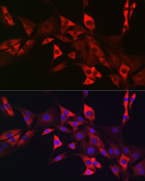
Immunofluorescence analysis of NIH/3T3 cells using CD117/c-Kit Rabbit pAb (CAB0357) at dilution of 1:50. Secondary antibody: Cy3-conjugated Goat anti-Rabbit IgG (H+L) (CABS007) at 1:500 dilution. Blue: DAPI for nuclear staining.