The KLHDC3 Antibody (CAB13741) is a high-quality antibody developed for reliable detection and analysis of target proteins. This antibody is produced in rabbits and has been validated for use in Western blot applications, providing reliable and accurate detection of the KLHDC3 protein in human samples.KLHDC3, a Kelch domain-containing protein, is known to interact with other proteins involved in ubiquitin-mediated protein degradation, suggesting a role in maintaining protein homeostasis within cells. Its function in the cell cycle and potential implications in disease make it an intriguing target for further investigation in research areas such as cancer biology, cell signaling, and molecular biology.
This antibody is validated for use in WB, IF/ICC, ELISA applications and has demonstrated reactivity against Human, Mouse, Rat samples.
Product Name:
KLHDC3 Antibody
SKU:
CAB13741
Size:
20μL, 100μL
Reactivity:
Human, Mouse, Rat
Conjugate:
Unconjugated
Immunogen:
Recombinant protein (or fragment).This information is considered to be commercially sensitive.
Recommended starting concentration is 1 μg/mL. Please optimize the concentration based on your specific assay requirements.
Synonyms:
PEAS, dJ20C7.3, KLHDC3
Positive Sample:
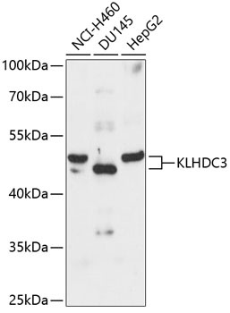
NCI-H460, DU145, HepG2
Cellular Localization:
Cytoplasm.
Calculated MW:
43kDa
Observed MW:
43kDa
The protein encoded by this gene contains six repeated kelch motifs that are structurally similar to recombination activating gene 2, a protein involved in the activation of the V(D)J recombination. In mouse, this gene is found to be expressed specifically in testis. Its expression in pachytene spermatocytes is localized to cytoplasma and meiotic chromatin, suggesting that this gene may be involved in meiotic recombination.
Purification Method
Affinity purification
Gene ID
116138
RRID
AB_2760601
Buffer Information
Store at -20℃. Avoid freeze / thaw cycles. Buffer: PBS containing 50% glycerol, preserved with proclin300 or sodium azide, pH 7.3.
Western blot analysis of various lysates using KLHDC3 Rabbit pAb (CAB13741) at 1:3000 dilution. Secondary antibody: HRP-conjugated Goat anti-Rabbit IgG (H+L) (CABS014) at 1:10000 dilution. Lysates/proteins: 25μg per lane. Blocking buffer: 3% nonfat dry milk in TBST. Detection: ECL Basic Kit (AbGn00020). Exposure time: 90s.
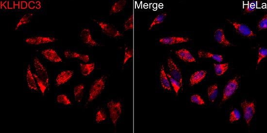
Immunofluorescence analysis of HeLa cells using KLHDC3 Rabbit pAb (CAB13741) at dilution of 1:100 (40x lens). Secondary antibody: Cy3-conjugated Goat anti-Rabbit IgG (H+L) (CABS007) at 1:500 dilution. Blue: DAPI for nuclear staining.