The [KO Validated] beta-Catenin Monoclonal Antibody (CAB19657) is a high-quality antibody developed for reliable detection and analysis of target proteins. The protein encoded by this gene is part of a complex of proteins that constitute adherens junctions (AJs). AJs are necessary for the creation and maintenance of epithelial cell layers by regulating cell growth and adhesion between cells. The encoded protein also anchors the actin cytoskeleton and may be responsible for transmitting the contact inhibition signal that causes cells to stop dividing once the epithelial sheet is complete. Finally, this protein binds to the product of the APC gene, which is mutated in adenomatous polyposis of the colon. Mutations in this gene are a cause of colorectal cancer (CRC), pilomatrixoma (PTR), medulloblastoma (MDB), and ovarian cancer. Alternative splicing results in multiple transcript variants.
This antibody is validated for use in WB, IHC-P, IP, ELISA, IF-P applications and has demonstrated reactivity against Human, Mouse, Rat samples.
Product Name:
[KO Validated] beta-Catenin Monoclonal Antibody
SKU:
CAB19657
Size:
100μL, 20μL
Reactivity:
Human, Mouse, Rat
Clone Number:
ARC0136
Conjugate:
Unconjugated
Immunogen:
Synthetic peptide. This information is considered to be commercially sensitive.
Tested Applications:
WBIHC-PIPELISAIF-P
Recommended Dilution:
WB
1:4000 - 1:20000
IP
0.5μg-4μg antibody for 400μg-600μg extracts of whole cells
IF-P
1:50 - 1:200
IHC-P
1:500 - 1:2000
ELISA
Recommended starting concentration is 1 μg/mL. Please optimize the concentration based on your specific assay requirements.
Synonyms:
EVR7, CTNNB, MRD19, NEDSDV, armadillo, in
Positive Sample:
HeLa, 293T, COS-7, Mouse brain
Cellular Localization:
Cell Junction, Cell Membrane, Cytoplasm, Nucleus, Adherens Junction, Centrosome, Cytoskeleton, Microtubule Organizing Center, Spindle Pole.
Calculated MW:
85kDa
Observed MW:
92kDa
The protein encoded by this gene is part of a complex of proteins that constitute adherens junctions (AJs). AJs are necessary for the creation and maintenance of epithelial cell layers by regulating cell growth and adhesion between cells. The encoded protein also anchors the actin cytoskeleton and may be responsible for transmitting the contact inhibition signal that causes cells to stop dividing once the epithelial sheet is complete. Finally, this protein binds to the product of the APC gene, which is mutated in adenomatous polyposis of the colon. Mutations in this gene are a cause of colorectal cancer (CRC), pilomatrixoma (PTR), medulloblastoma (MDB), and ovarian cancer. Alternative splicing results in multiple transcript variants.
Purification Method
Affinity purification
Gene ID
1499
RRID
AB_2862719
Buffer Information
Store at -20℃. Avoid freeze / thaw cycles. Buffer: PBS containing 50% glycerol and 0.05% BSA, preserved with proclin300 or sodium azide, pH 7.3.
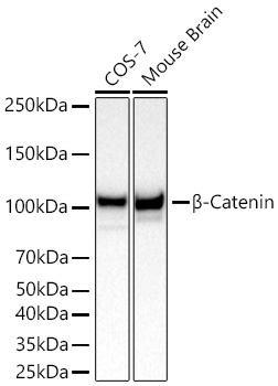
Western blot analysis of various lysates using [KO Validated] β-Catenin Rabbit mAb (CAB19657) at 1:4000 dilution incubated at room temperature for 1.5 hours. Secondary antibody: HRP-conjugated Goat anti-Rabbit IgG (H+L) (AS014) at 1:10000 dilution. Lysates/proteins: 25 μg per lane. Blocking buffer: 3% nonfat dry milk in TBST. Detection: ECL Basic Kit (AbGn00020). Exposure time: 5s.
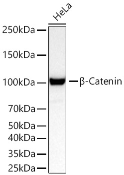
Western blot analysis of lysates from HeLa cells using [KO Validated] β-Catenin Rabbit mAb (CAB19657) at 1:4000 dilution incubated at room temperature for 1.5 hours. Secondary antibody: HRP-conjugated Goat anti-Rabbit IgG (H+L) (AS014) at 1:10000 dilution. Lysates/proteins: 25 μg per lane. Blocking buffer: 3% nonfat dry milk in TBST. Detection: ECL Basic Kit (AbGn00020). Exposure time: 45s.
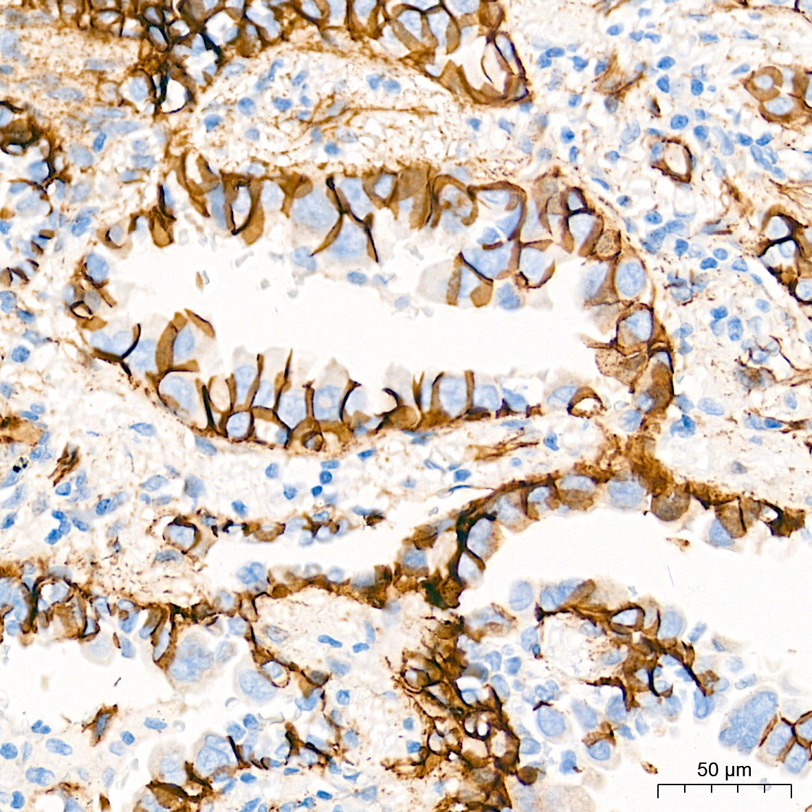
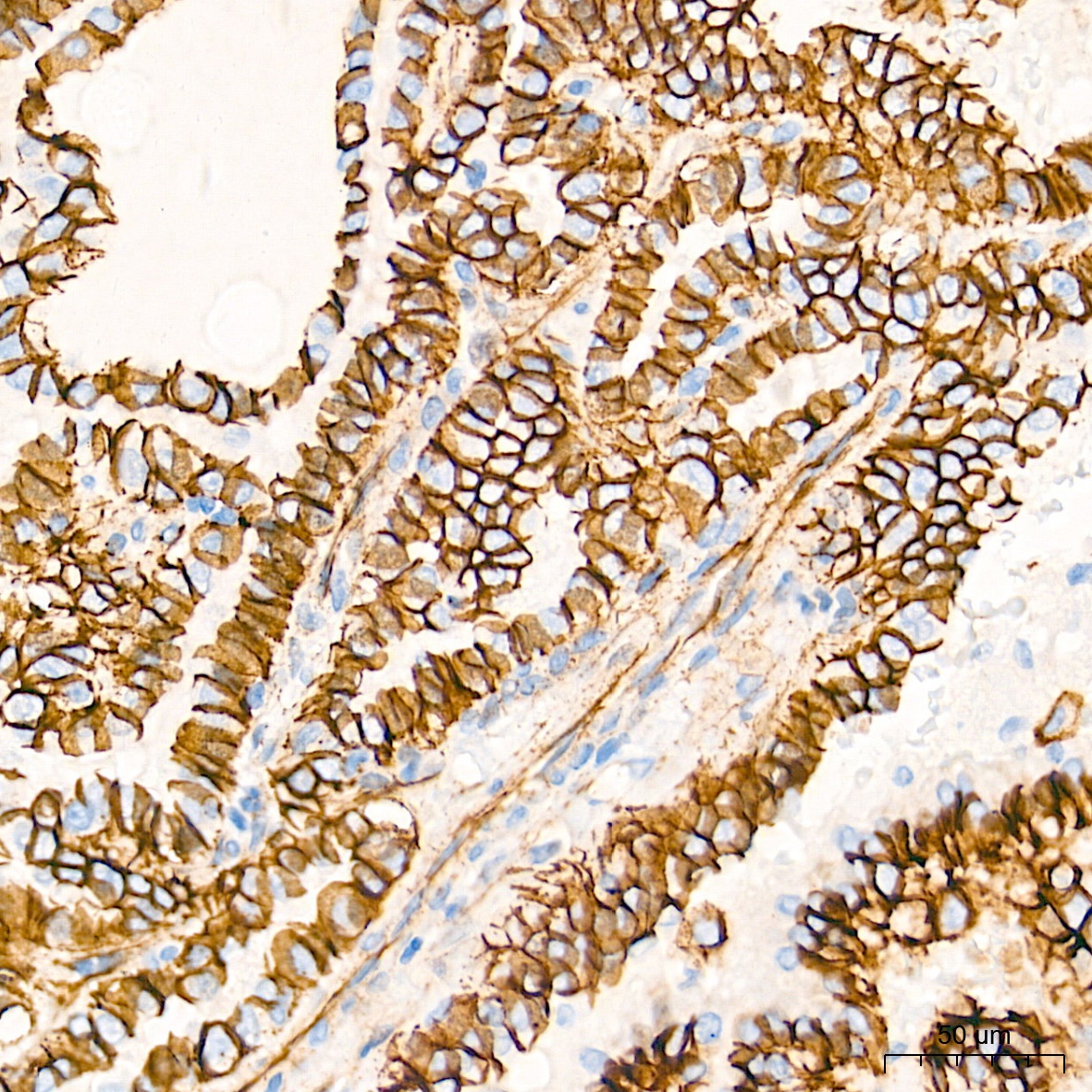
Immunohistochemistry analysis of paraffin-embedded Human lung cancer tissue using [KO Validated] β-Catenin Rabbit mAb (CAB19657) at a dilution of 1:800 (40x lens). High pressure antigen retrieval performed with 0.01M Tris-EDTA Buffer (pH 9.0) prior to IHC staining.
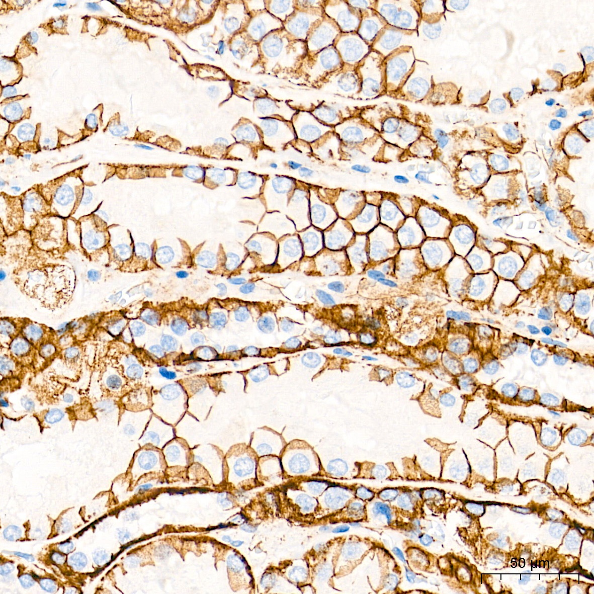
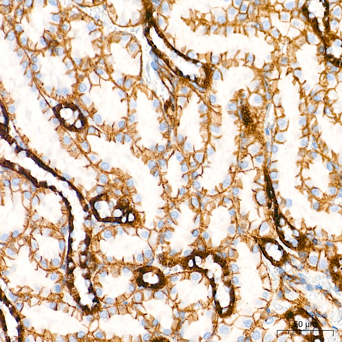
Immunohistochemistry analysis of paraffin-embedded Human kidney tissue using [KO Validated] β-Catenin Rabbit mAb (CAB19657) at a dilution of 1:800 (40x lens). High pressure antigen retrieval performed with 0.01M Tris-EDTA Buffer (pH 9.0) prior to IHC staining.
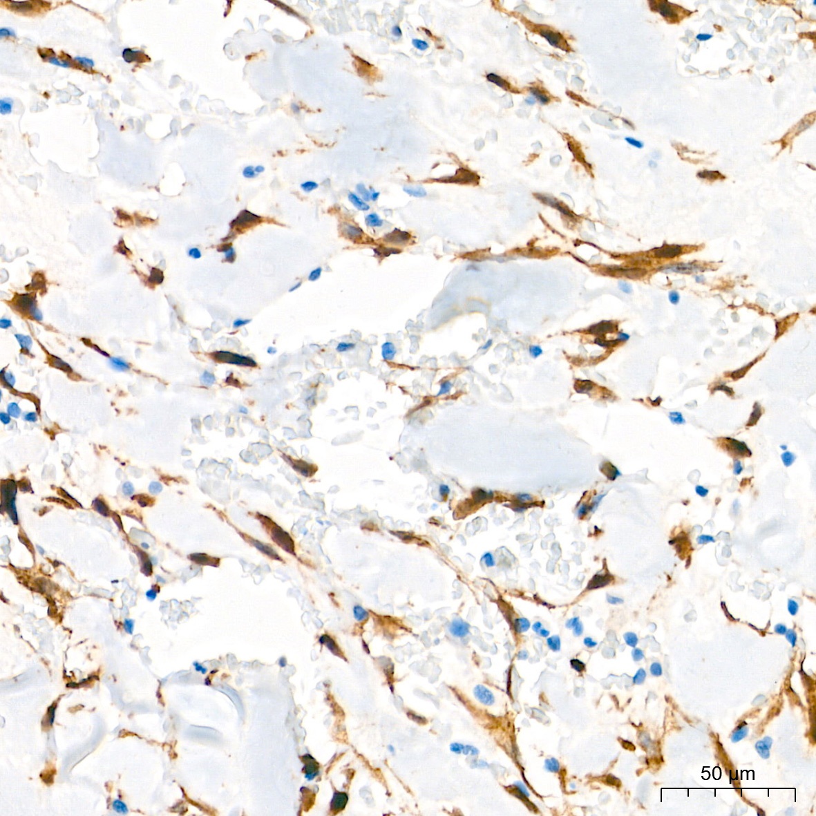
Immunohistochemistry analysis of paraffin-embedded Human solitary fibrous tumor tissue using [KO Validated] β-Catenin Rabbit mAb (CAB19657) at a dilution of 1:800 (40x lens). High pressure antigen retrieval performed with 0.01M Tris-EDTA Buffer (pH 9.0) prior to IHC staining.
Immunohistochemistry analysis of paraffin-embedded Human thyroid cancer tissue using [KO Validated] β-Catenin Rabbit mAb (CAB19657) at a dilution of 1:800 (40x lens). High pressure antigen retrieval performed with 0.01M Tris-EDTA Buffer (pH 9.0) prior to IHC staining.
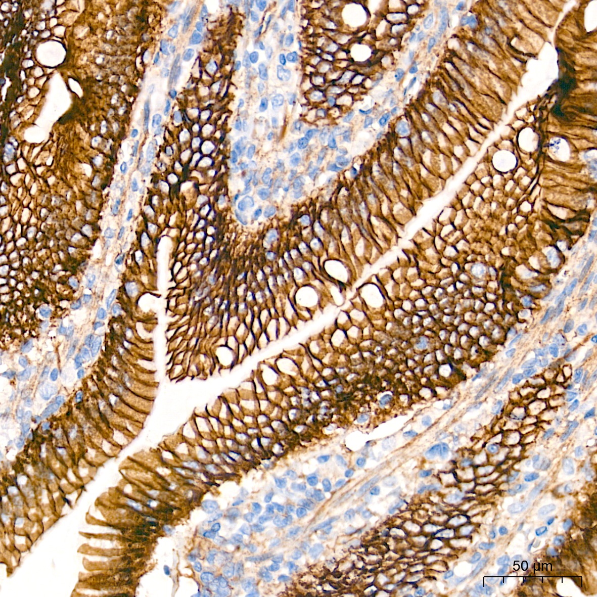
Immunohistochemistry analysis of paraffin-embedded Rat intestine tissue using [KO Validated] β-Catenin Rabbit mAb (CAB19657) at a dilution of 1:800 (40x lens). High pressure antigen retrieval performed with 0.01M Tris-EDTA Buffer (pH 9.0) prior to IHC staining.
Immunohistochemistry analysis of paraffin-embedded Rat kidney tissue using [KO Validated] β-Catenin Rabbit mAb (CAB19657) at a dilution of 1:800 (40x lens). High pressure antigen retrieval performed with 0.01M Tris-EDTA Buffer (pH 9.0) prior to IHC staining.
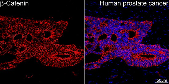
Confocal imaging of paraffin-embedded Human prostate cancer tissue using [KO Validated] β-Catenin Rabbit mAb (CAB19657, dilution 1:200) followed by a further incubation with Cy3 Goat Anti-Rabbit IgG (H+L) (AS007, dilution 1:500) (Red). DAPI was used for nuclear staining (Blue). High pressure antigen retrieval performed with 0.01M Citrate Buffer (pH 6.0) prior to IF staining. Objective: 40x.
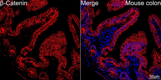
Confocal imaging of paraffin-embedded Mouse colon tissue using [KO Validated] β-Catenin Rabbit mAb (CAB19657, dilution 1:200) followed by a further incubation with Cy3 Goat Anti-Rabbit IgG (H+L) (AS007, dilution 1:500) (Red). DAPI was used for nuclear staining (Blue). High pressure antigen retrieval performed with 0.01M Citrate Buffer (pH 6.0) prior to IF staining. Objective: 40x.
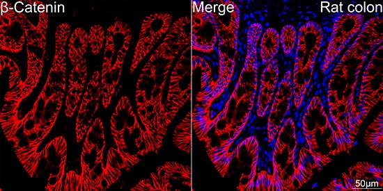
Confocal imaging of paraffin-embedded Rat colon tissue using [KO Validated] β-Catenin Rabbit mAb (CAB19657, dilution 1:200) followed by a further incubation with Cy3 Goat Anti-Rabbit IgG (H+L) (AS007, dilution 1:500) (Red). DAPI was used for nuclear staining (Blue). High pressure antigen retrieval performed with 0.01M Citrate Buffer (pH 6.0) prior to IF staining. Objective: 40x.