The [KO Validated] METTL3 Monoclonal Antibody (CAB19079) is a high-quality antibody developed for reliable detection and analysis of target proteins. This monoclonal antibody, created using rabbit-derived cells, is highly specific and sensitive, making it an ideal choice for applications such as Western blotting and immunohistochemistry.METTL3 plays a critical role in various cellular processes, including RNA metabolism, protein synthesis, and epigenetic regulation. Dysregulation of METTL3 has been linked to numerous diseases, including cancer, neurological disorders, and metabolic diseases.
This antibody is validated for use in WB, IHC-P, IF/ICC, IP, ELISA applications and has demonstrated reactivity against Human, Mouse, Rat samples.
Product Name:
[KO Validated] METTL3 Monoclonal Antibody
SKU:
CAB19079
Size:
20μL, 100μL
Reactivity:
Human, Mouse, Rat
Clone Number:
ARC0487
Conjugate:
Unconjugated
Immunogen:
Recombinant protein (or fragment).This information is considered to be commercially sensitive.
0.5μg-4μg antibody for 200μg-400μg extracts of whole cells
IF/ICC
1:100 - 1:400
IHC-P
1:200 - 1:800
ELISA
Recommended starting concentration is 1 μg/mL. Please optimize the concentration based on your specific assay requirements.
Synonyms:
M6A, IME4, Spo8, MT-A70, hMETTL3, METTL3
Positive Sample:
HeLa, 293T, 293F, Mouse brain, Rat brain
Cellular Localization:
Nucleus Speckle.
Calculated MW:
64kDa
Observed MW:
75kDa
This gene encodes the 70 kDa subunit of MT-A which is part of N6-adenosine-methyltransferase. This enzyme is involved in the posttranscriptional methylation of internal adenosine residues in eukaryotic mRNAs, forming N6-methyladenosine.
Purification Method
Affinity purification
Gene ID
56339
RRID
AB_2862571
Buffer Information
Store at -20℃. Avoid freeze / thaw cycles. Buffer: PBS containing 50% glycerol and 0.05% BSA, preserved with proclin300 or sodium azide, pH 7.3.
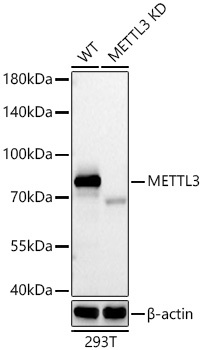
Western blot analysis of lysates from wild type (WT) and METTL3 knockdown (KD) 293T cells using METTL3 Rabbit mAb (CAB19079) at 1:1000 dilution incubated overnight at 4℃. Secondary antibody: HRP-conjugated Goat anti-Rabbit IgG (H+L) (CABS014) at 1:10000 dilution. Lysates/proteins: 25 μg per lane. Blocking buffer: 3% nonfat dry milk in TBST. Detection: ECL Basic Kit (AbGn00020). Exposure time: 20s.
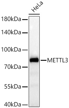
Western blot analysis of lysates from HeLa cells using [KD Validated] METTL3 Rabbit mAb (CAB19079) at 1:1000 dilution incubated overnight at 4℃. Secondary antibody: HRP-conjugated Goat anti-Rabbit IgG (H+L) (CABS014) at 1:10000 dilution. Lysates/proteins: 25 μg per lane. Blocking buffer: 3% nonfat dry milk in TBST. Detection: ECL Basic Kit (AbGn00020). Exposure time: 20s.
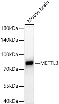
Western blot analysis of lysates from Mouse brain using [KD Validated] METTL3 Rabbit mAb (CAB19079) at 1:1000 dilution incubated overnight at 4℃. Secondary antibody: HRP-conjugated Goat anti-Rabbit IgG (H+L) (CABS014) at 1:10000 dilution. Lysates/proteins: 25 μg per lane. Blocking buffer: 3% nonfat dry milk in TBST. Detection: ECL Basic Kit (AbGn00020). Exposure time: 60s.
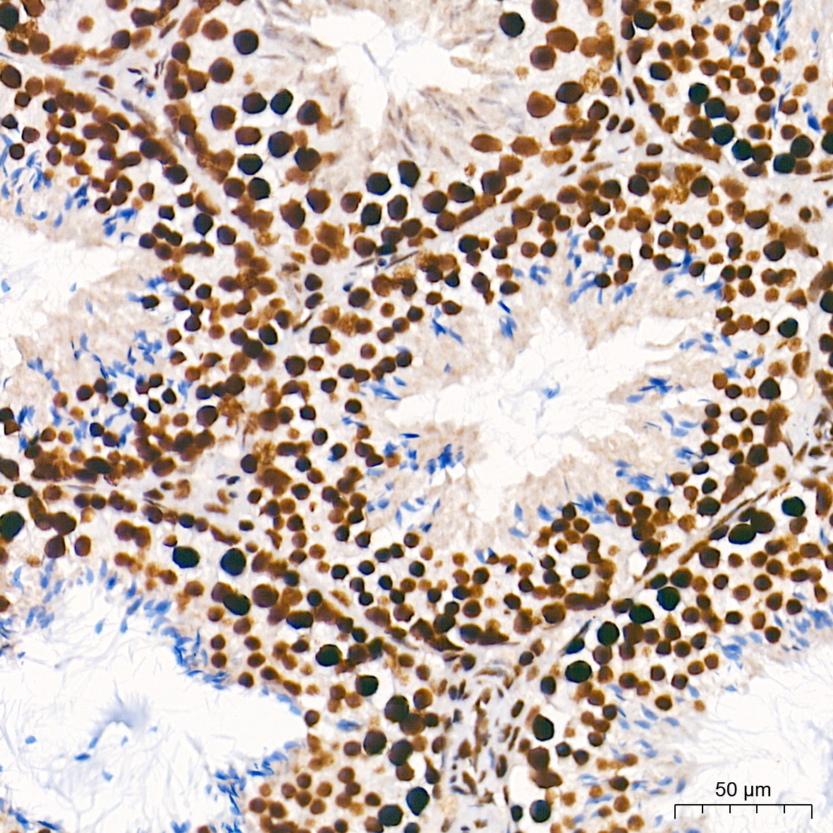
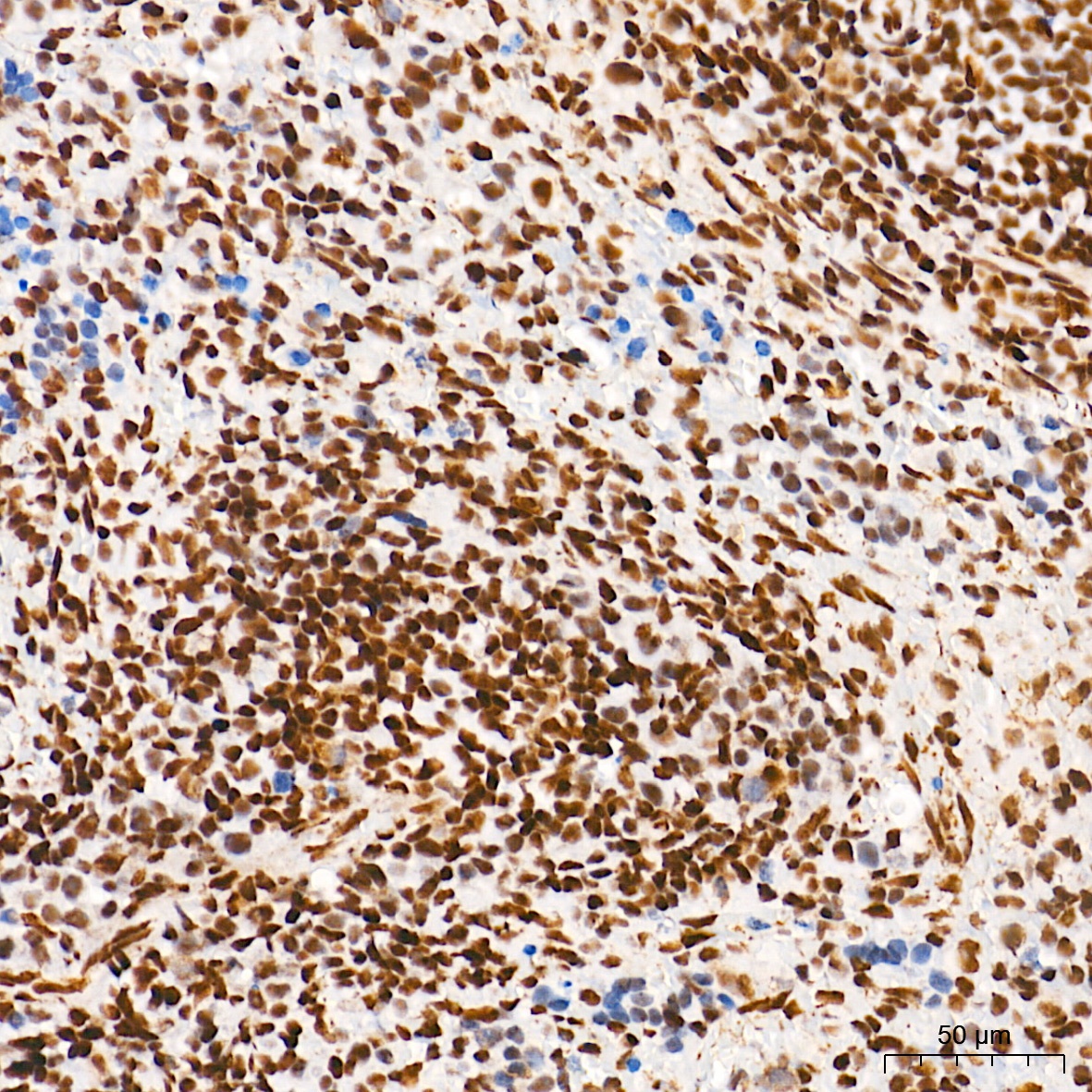
Immunohistochemistry analysis of paraffin-embedded Mouse testis tissue using [KD Validated] METTL3 Rabbit mAb (CAB19079) at a dilution of 1:200 (40x lens). High pressure antigen retrieval performed with 0.01M Tris-EDTA Buffer (pH 9.0) prior to IHC staining.
Immunohistochemistry analysis of paraffin-embedded Rat spleen tissue using [KD Validated] METTL3 Rabbit mAb (CAB19079) at a dilution of 1:200 (40x lens). High pressure antigen retrieval performed with 0.01M Tris-EDTA Buffer (pH 9.0) prior to IHC staining.
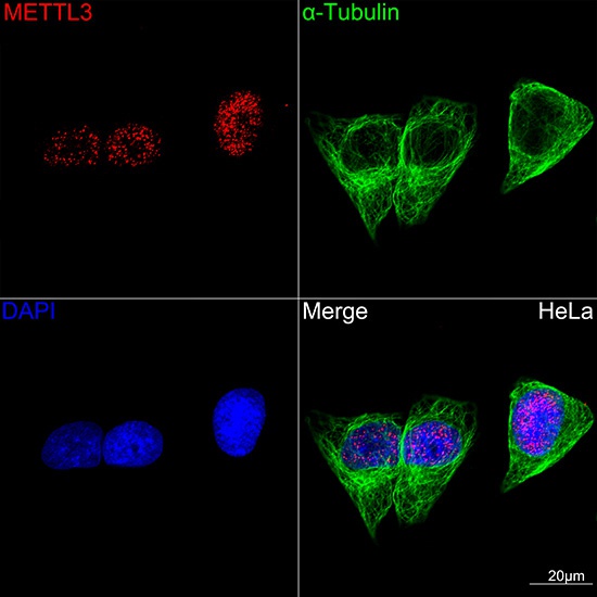
Confocal imaging of HeLa cells using [KD Validated] METTL3 Rabbit mAb (CAB19079,dilution 1:100)(Red) followed by a further incubation with Cy3-conjugated Goat Anti-Rabbit IgG (H+L) (CABS007, dilution 1:500) (Red). The cells were counterstained with α-Tubulin mAb (AC012, dilution 1:400) followed by incubation with ABflo® 488-conjugated Goat Anti-Mouse IgG (H+L) (CABS076, dilution 1:500) (Green). DAPI was used for nuclear staining (blue). Objective: 100x.