The [KO Validated] METTL3 Monoclonal Antibody (CAB19079) is a high-quality antibody developed for reliable detection and analysis of target proteins. This gene encodes the 70 kDa subunit of MT-A which is part of N6-adenosine-methyltransferase. This enzyme is involved in the posttranscriptional methylation of internal adenosine residues in eukaryotic mRNAs, forming N6-methyladenosine.
This antibody is validated for use in WB, IHC-P, IF/ICC, IP, ELISA applications and has demonstrated reactivity against Human, Mouse, Rat samples.
Product Name:
[KO Validated] METTL3 Monoclonal Antibody
SKU:
CAB19079
Size:
100μL, 20μL
Reactivity:
Human, Mouse, Rat
Clone Number:
ARC0487
Conjugate:
Unconjugated
Immunogen:
Recombinant protein (or fragment).This information is considered to be commercially sensitive.
Tested Applications:
WBIHC-PIF/ICCIPELISA
Recommended Dilution:
WB
1:1000 - 1:2000
IP
0.5μg-4μg antibody for 200μg-400μg extracts of whole cells
IF/ICC
1:100 - 1:400
IHC-P
1:200 - 1:800
ELISA
Recommended starting concentration is 1 μg/mL. Please optimize the concentration based on your specific assay requirements.
Synonyms:
M6A, IME4, Spo8, MT-A70, hMETTL3, METTL3
Positive Sample:
HeLa, 293T, 293F, Mouse brain, Rat brain
Cellular Localization:
Nucleus Speckle.
Calculated MW:
64kDa
Observed MW:
75kDa
This gene encodes the 70 kDa subunit of MT-A which is part of N6-adenosine-methyltransferase. This enzyme is involved in the posttranscriptional methylation of internal adenosine residues in eukaryotic mRNAs, forming N6-methyladenosine.
Purification Method
Affinity purification
Gene ID
56339
RRID
AB_2862571
Buffer Information
Store at -20℃. Avoid freeze / thaw cycles. Buffer: PBS containing 50% glycerol and 0.05% BSA, preserved with proclin300 or sodium azide, pH 7.3.
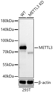
Western blot analysis of lysates from wild type (WT) and METTL3 knockdown (KD) 293T cells using METTL3 Rabbit mAb (CAB19079) at 1:1000 dilution incubated overnight at 4℃. Secondary antibody: HRP-conjugated Goat anti-Rabbit IgG (H+L) (AS014) at 1:10000 dilution. Lysates/proteins: 25 μg per lane. Blocking buffer: 3% nonfat dry milk in TBST. Detection: ECL Basic Kit (AbGn00020). Exposure time: 20s.
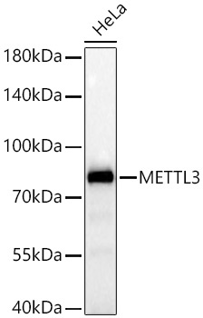
Western blot analysis of lysates from HeLa cells using [KD Validated] METTL3 Rabbit mAb (CAB19079) at 1:1000 dilution incubated overnight at 4℃. Secondary antibody: HRP-conjugated Goat anti-Rabbit IgG (H+L) (AS014) at 1:10000 dilution. Lysates/proteins: 25 μg per lane. Blocking buffer: 3% nonfat dry milk in TBST. Detection: ECL Basic Kit (AbGn00020). Exposure time: 20s.
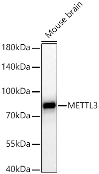
Western blot analysis of lysates from Mouse brain using [KD Validated] METTL3 Rabbit mAb (CAB19079) at 1:1000 dilution incubated overnight at 4℃. Secondary antibody: HRP-conjugated Goat anti-Rabbit IgG (H+L) (AS014) at 1:10000 dilution. Lysates/proteins: 25 μg per lane. Blocking buffer: 3% nonfat dry milk in TBST. Detection: ECL Basic Kit (AbGn00020). Exposure time: 60s.
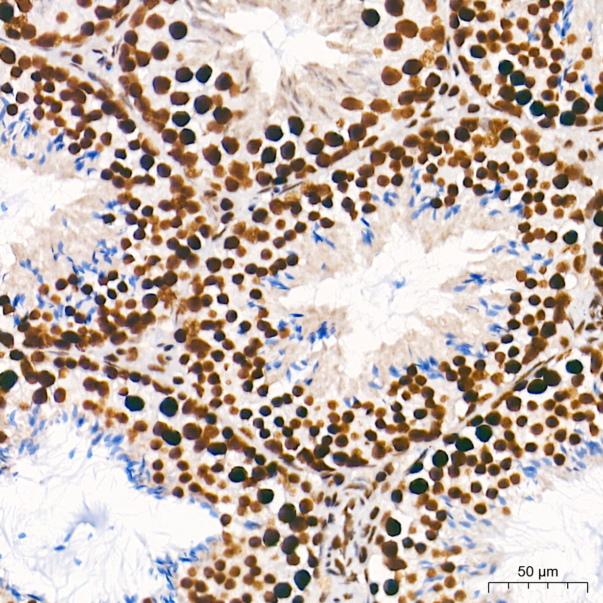
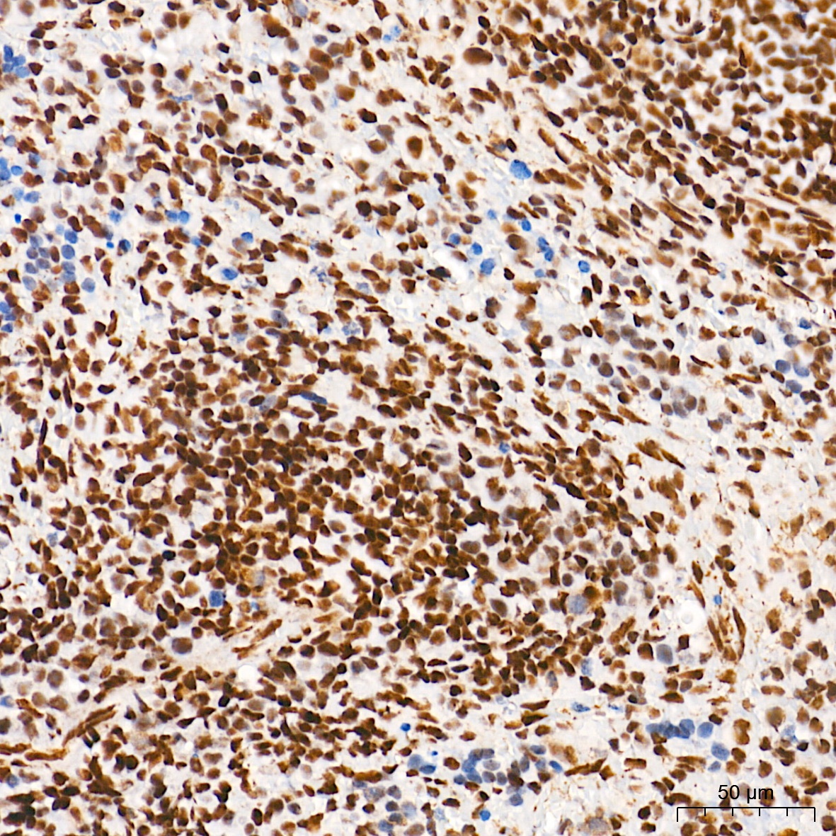
Immunohistochemistry analysis of paraffin-embedded Mouse testis tissue using [KD Validated] METTL3 Rabbit mAb (CAB19079) at a dilution of 1:200 (40x lens). High pressure antigen retrieval performed with 0.01M Tris-EDTA Buffer (pH 9.0) prior to IHC staining.
Immunohistochemistry analysis of paraffin-embedded Rat spleen tissue using [KD Validated] METTL3 Rabbit mAb (CAB19079) at a dilution of 1:200 (40x lens). High pressure antigen retrieval performed with 0.01M Tris-EDTA Buffer (pH 9.0) prior to IHC staining.
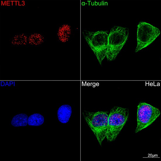
Confocal imaging of HeLa cells using [KD Validated] METTL3 Rabbit mAb (CAB19079,dilution 1:100)(Red) followed by a further incubation with Cy3-conjugated Goat Anti-Rabbit IgG (H+L) (AS007, dilution 1:500) (Red). The cells were counterstained with α-Tubulin mAb (AC012, dilution 1:400) followed by incubation with ABflo® 488-conjugated Goat Anti-Mouse IgG (H+L) (AS076, dilution 1:500) (Green). DAPI was used for nuclear staining (blue). Objective: 100x.