The [KO Validated] NCOA2 Antibody (CAB10280) is a high-quality antibody developed for reliable detection and analysis of target proteins. The antibody, raised in rabbits, is highly reactive with human samples and is validated for use in Western blot applications. It binds specifically to the NCOA2 protein, allowing for accurate detection and analysis in various cell types, making it ideal for studies in molecular biology and cancer research.
This antibody is validated for use in WB, IHC-P, ELISA applications and has demonstrated reactivity against Human, Rat samples.
Product Name:
[KO Validated] NCOA2 Antibody
SKU:
CAB10280
Size:
20μL, 100μL
Reactivity:
Human, Rat
Conjugate:
Unconjugated
Immunogen:
Recombinant protein (or fragment).This information is considered to be commercially sensitive.
Recommended starting concentration is 1 μg/mL. Please optimize the concentration based on your specific assay requirements.
Synonyms:
SRC2, TIF2, GRIP1, KAT13C, NCoA-2, bHLHe75, A2
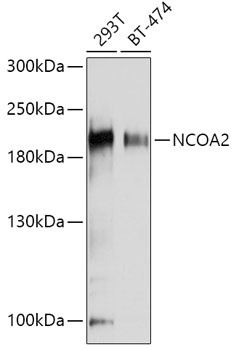
Positive Sample:
293T, BT-474
Cellular Localization:
Nucleus.
Calculated MW:
159kDa
Observed MW:
200kDa
The protein encoded by this gene functions as a transcriptional coactivator for nuclear hormone receptors, including steroid, thyroid, retinoid, and vitamin D receptors. The encoded protein acts as an intermediary factor for the ligand-dependent activity of these nuclear receptors, which regulate their target genes upon binding of cognate response elements. This gene has been found to be involved in translocations that result in fusions with other genes in various cancers, including the lysine acetyltransferase 6A (KAT6A) gene in acute myeloid leukemia, the ETS variant 6 (ETV6) gene in acute lymphoblastic leukemia, and the hes related family bHLH transcription factor with YRPW motif 1 (HEY1) gene in mesenchymal chondrosarcoma. Alternative splicing results in multiple transcript variants.
Purification Method
Affinity purification
Gene ID
10499
RRID
AB_2757809
Buffer Information
Store at -20℃. Avoid freeze / thaw cycles. Buffer: PBS containing 50% glycerol, preserved with proclin300 or sodium azide, pH 7.3.
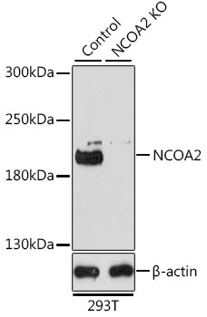
Western blot analysis of lysates from wild type (WT) and NCOA2 knockout (KO) 293T cells, using [KO Validated] NCOA2 Rabbit pAb (CAB10280) at 1:1000 dilution. Secondary antibody: HRP-conjugated Goat anti-Rabbit IgG (H+L) (CABS014) at 1:10000 dilution. Lysates/proteins: 25μg per lane. Blocking buffer: 3% nonfat dry milk in TBST. Detection: ECL Basic Kit (AbGn00020). Exposure time: 3min.
Western blot analysis of various lysates using [KO Validated] NCOA2 Rabbit pAb (CAB10280) at 1:1000 dilution. Secondary antibody: HRP-conjugated Goat anti-Rabbit IgG (H+L) (CABS014) at 1:10000 dilution. Lysates/proteins: 25μg per lane. Blocking buffer: 3% nonfat dry milk in TBST. Detection: ECL Basic Kit (AbGn00020). Exposure time: 180s.
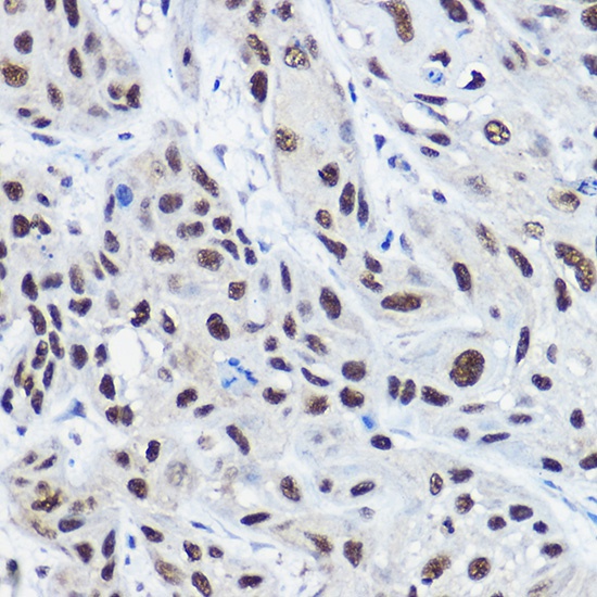
Immunohistochemistry analysis of paraffin-embedded Human esophageal cancer using [KO Validated] NCOA2 Rabbit pAb (CAB10280) at dilution of 1:100 (40x lens). High pressure antigen retrieval performed with 0.01M Citrate buffer (pH 6.0) prior to IHC staining.