The [KO Validated] NDUFS3 Antibody (CAB8013) is a high-quality antibody developed for reliable detection and analysis of target proteins. This antibody, generated in rabbits, is highly specific to human samples and has been validated for use in Western blot applications. With its ability to bind specifically to the NDUFS3 protein, researchers can accurately detect and analyze this key component of mitochondrial function in various cell types.NDUFS3 is essential for the function of complex I in the electron transport chain, making it crucial for cellular energy production.
This antibody is validated for use in WB, IP, ELISA applications and has demonstrated reactivity against Human, Mouse, Rat samples.
Product Name:
[KO Validated] NDUFS3 Antibody
SKU:
CAB8013
Size:
20μL, 100μL
Reactivity:
Human, Mouse, Rat
Conjugate:
Unconjugated
Immunogen:
Recombinant protein (or fragment).This information is considered to be commercially sensitive.
0.5μg-4μg antibody for 400μg-600μg extracts of whole cells
ELISA
Recommended starting concentration is 1 μg/mL. Please optimize the concentration based on your specific assay requirements.
Synonyms:
CI-30, MC1DN8, NDUFS3
Positive Sample:
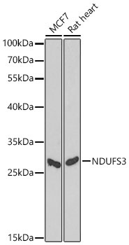
HepG2, Mouse kidney, Mouse heart, Mouse skeletal muscle, Rat kidney, Rat heart, Rat skeletal muscle, MCF7, Rat heart
Cellular Localization:
Mitochondrion Inner Membrane.
Calculated MW:
30kDa
Observed MW:
26kDa/30kDa
This gene encodes one of the iron-sulfur protein (IP) components of mitochondrial NADH:ubiquinone oxidoreductase (complex I). Mutations in this gene are associated with Leigh syndrome resulting from mitochondrial complex I deficiency.
Purification Method
Affinity purification
Gene ID
4722
RRID
AB_2863565
Buffer Information
Store at -20℃. Avoid freeze / thaw cycles. Buffer: PBS containing 50% glycerol, preserved with proclin300 or sodium azide, pH 7.3.
Western blot analysis of various lysates using NDUFS3 Rabbit pAb (CAB8013) at 1:1000 dilution. Secondary antibody: HRP-conjugated Goat anti-Rabbit IgG (H+L) (CABS014) at 1:10000 dilution. Lysates / proteins: 25 μg per lane. Blocking buffer: 3 % nonfat dry milk in TBST. Detection: ECL Basic Kit (AbGn00020). Exposure time: 90s.
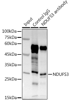
Immunoprecipitation analysis of 600 μg extracts of Mouse kidney using 3 μg NDUFS3 antibody (CAB8013). Western blot was performed from the immunoprecipitate using NDUFS3 antibody (CAB8013) at a dilution of 1:1000.