The [KO Validated] S100A11 Antibody (CAB5486) is a high-quality antibody developed for reliable detection and analysis of target proteins. This antibody, produced in rabbits, is highly specific for human samples and has been validated for use in Western blotting applications. By binding to the S100 A11 protein, this antibody enables the detection and analysis of S100 A11 in various cell types, making it a versatile tool for studies in cell biology, cancer research, and inflammation.
This antibody is validated for use in WB, IF/ICC, IP, ELISA applications and has demonstrated reactivity against Human samples.
Product Name:
[KO Validated] S100A11 Antibody
SKU:
CAB5486
Size:
20μL, 100μL
Reactivity:
Human
Conjugate:
Unconjugated
Immunogen:
Recombinant protein (or fragment).This information is considered to be commercially sensitive.
0.5μg-4μg antibody for 200μg-400μg extracts of whole cells
ELISA
Recommended starting concentration is 1 μg/mL. Please optimize the concentration based on your specific assay requirements.
Synonyms:
MLN70, S100C, HEL-S-43, 11
Positive Sample:
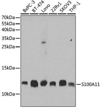
BxPC-3, BT-474, LOVO, 22Rv1, SKOV3, THP-1
Cellular Localization:
Cytoplasm, Nucleus.
Calculated MW:
12kDa
Observed MW:
12kDa
The protein encoded by this gene is a member of the S100 family of proteins containing 2 EF-hand calcium-binding motifs. S100 proteins are localized in the cytoplasm and/or nucleus of a wide range of cells, and involved in the regulation of a number of cellular processes such as cell cycle progression and differentiation. S100 genes include at least 13 members which are located as a cluster on chromosome 1q21. This protein may function in motility, invasion, and tubulin polymerization. Chromosomal rearrangements and altered expression of this gene have been implicated in tumor metastasis.
Purification Method
Affinity purification
Gene ID
6282
RRID
AB_2863504
Buffer Information
Store at -20℃. Avoid freeze / thaw cycles. Buffer: PBS containing 50% glycerol, preserved with proclin300 or sodium azide, pH 7.3.
Western blot analysis of various lysates using [KO Validated] S100A11 Rabbit pAb (CAB5486) at 1:1000 dilution. Secondary antibody: HRP-conjugated Goat anti-Rabbit IgG (H+L) (CABS014) at 1:10000 dilution. Lysates/proteins: 25μg per lane. Blocking buffer: 3% nonfat dry milk in TBST. Detection: ECL Basic Kit (AbGn00020). Exposure time: 10s.
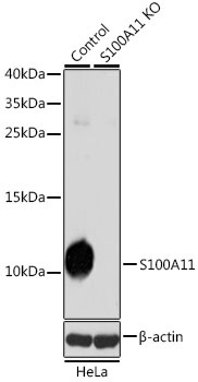
Western blot analysis of lysates from wild type (WT) and S100A11 knockout (KO) HeLa cells, using [KO Validated] S100A11 Rabbit pAb (CAB5486) at 1:1000 dilution. Secondary antibody: HRP-conjugated Goat anti-Rabbit IgG (H+L) (CABS014) at 1:10000 dilution. Lysates/proteins: 25μg per lane. Blocking buffer: 3% nonfat dry milk in TBST. Detection: ECL Basic Kit (AbGn00020). Exposure time: 10s.
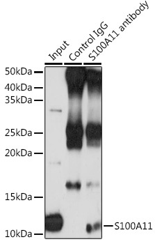
Immunoprecipitation analysis of 200 μg extracts of THP-1 cells using 3 μg S100A11 antibody (CAB5486). Western blot was performed from the immunoprecipitate using S100A11 antibody (CAB5486) at a dilution of 1:1000.