The [KO Validated] Smad4 Monoclonal Antibody (CAB19116) is a high-quality antibody developed for reliable detection and analysis of target proteins. This gene encodes a member of the Smad family of signal transduction proteins. Smad proteins are phosphorylated and activated by transmembrane serine-threonine receptor kinases in response to transforming growth factor (TGF)-beta signaling. The product of this gene forms homomeric complexes and heteromeric complexes with other activated Smad proteins, which then accumulate in the nucleus and regulate the transcription of target genes. This protein binds to DNA and recognizes an 8-bp palindromic sequence (GTCTAGAC) called the Smad-binding element (SBE). The protein acts as a tumor suppressor and inhibits epithelial cell proliferation. It may also have an inhibitory effect on tumors by reducing angiogenesis and increasing blood vessel hyperpermeability. The encoded protein is a crucial component of the bone morphogenetic protein signaling pathway. The Smad proteins are subject to complex regulation by post-translational modifications. Mutations or deletions in this gene have been shown to result in pancreatic cancer, juvenile polyposis syndrome, and hereditary hemorrhagic telangiectasia syndrome.
This antibody is validated for use in WB, IHC-P, IP, ChIP, ELISA, CUT&Tag applications and has demonstrated reactivity against Human, Mouse, Rat samples.
Product Name:
[KO Validated] Smad4 Monoclonal Antibody
SKU:
CAB19116
Size:
100μL, 20μL
Reactivity:
Human, Mouse, Rat
Clone Number:
ARC5009-06
Conjugate:
Unconjugated
Immunogen:
Recombinant protein (or fragment).This information is considered to be commercially sensitive.
Tested Applications:
WBIHC-PIPChIPELISACUT&Tag
Recommended Dilution:
WB
1:1000 - 1:6000
IHC-P
1:500 - 1:2000
IP
0.5μg-4μg antibody for 200μg-400μg extracts of whole cells
ELISA
Recommended starting concentration is 1 μg/mL. Please optimize the concentration based on your specific assay requirements.
ChIP
5μg antibody for 10μg-15μg of Chromatin
CUT&Tag
10⁵ cells /1 μg
Synonyms:
JIP, DPC4, MADH4, MYHRS, d4
Positive Sample:
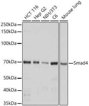
293T, HCT 116, HepG2, C6, NIH/3T3, Mouse lung
Cellular Localization:
Cytoplasm, Nucleus.
Calculated MW:
60kDa
Observed MW:
70kDa
This gene encodes a member of the Smad family of signal transduction proteins. Smad proteins are phosphorylated and activated by transmembrane serine-threonine receptor kinases in response to transforming growth factor (TGF)-beta signaling. The product of this gene forms homomeric complexes and heteromeric complexes with other activated Smad proteins, which then accumulate in the nucleus and regulate the transcription of target genes. This protein binds to DNA and recognizes an 8-bp palindromic sequence (GTCTAGAC) called the Smad-binding element (SBE). The protein acts as a tumor suppressor and inhibits epithelial cell proliferation. It may also have an inhibitory effect on tumors by reducing angiogenesis and increasing blood vessel hyperpermeability. The encoded protein is a crucial component of the bone morphogenetic protein signaling pathway. The Smad proteins are subject to complex regulation by post-translational modifications. Mutations or deletions in this gene have been shown to result in pancreatic cancer, juvenile polyposis syndrome, and hereditary hemorrhagic telangiectasia syndrome.
Purification Method
Affinity purification
Gene ID
4089
RRID
AB_2862609
Buffer Information
Store at -20℃. Avoid freeze / thaw cycles. Buffer: PBS containing 50% glycerol and 0.05% BSA, preserved with proclin300 or sodium azide, pH 7.3.
Western blot analysis of various lysates using [KO Validated] Smad4 Rabbit mAb (CAB19116) at 1:1000 dilution incubated overnight at 4℃. Secondary antibody: HRP-conjugated Goat anti-Rabbit IgG (H+L) (AS014) at 1:10000 dilution. Lysates/proteins: 25 μg per lane. Blocking buffer: 3% nonfat dry milk in TBST. Detection: ECL Basic Kit (AbGn00020). Exposure time: 3s.
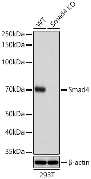
Western blot analysis of lysates from wild type (WT) and Smad4 knockout (KO) 293T cells using [KO Validated] Smad4 Rabbit mAb (CAB19116) at 1:1000 dilution incubated overnight at 4℃. Secondary antibody: HRP-conjugated Goat anti-Rabbit IgG (H+L) (AS014) at 1:10000 dilution. Lysates/proteins: 25 μg per lane. Blocking buffer: 3% nonfat dry milk in TBST. Detection: ECL Basic Kit (AbGn00020). Exposure time: 3s.
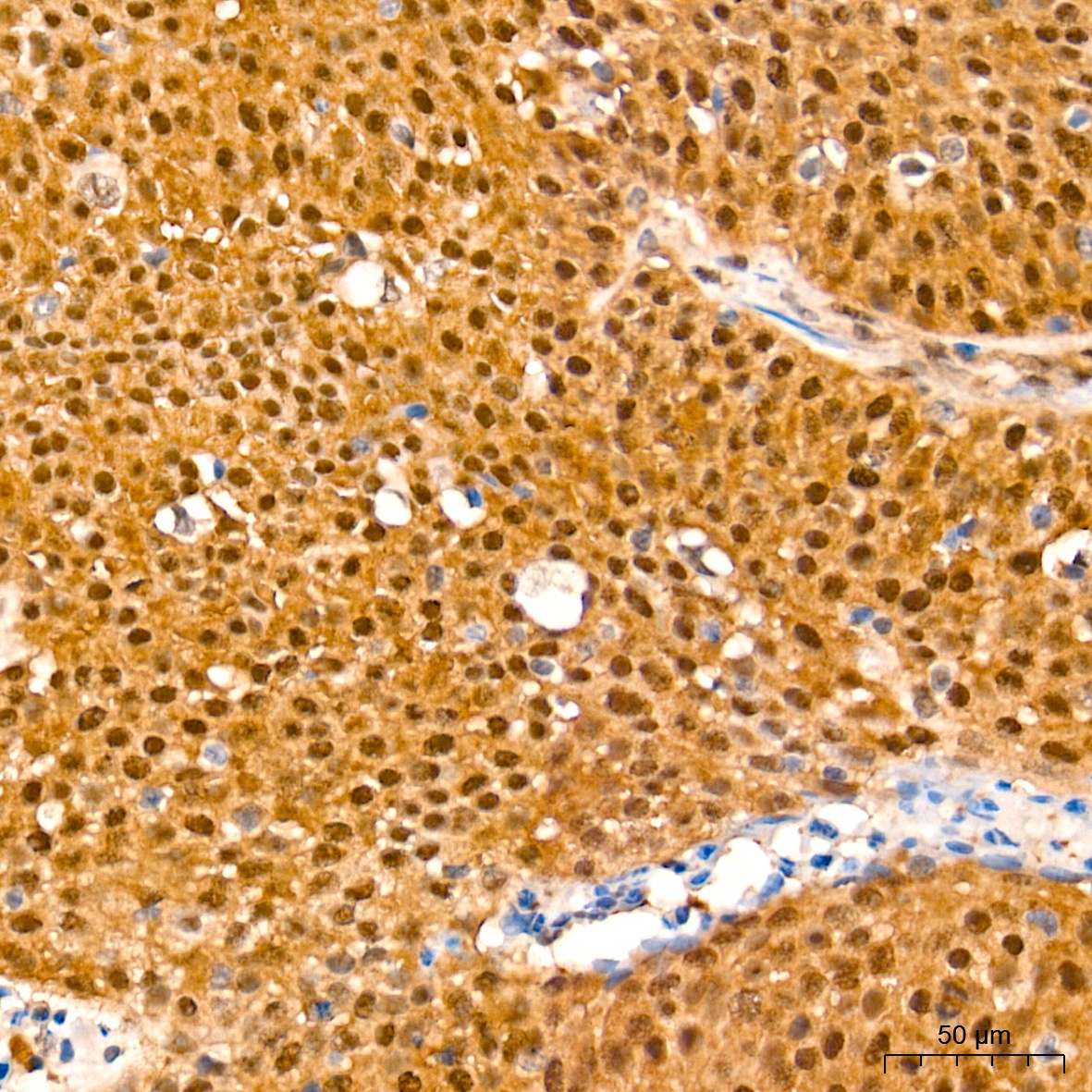
Immunohistochemistry analysis of paraffin-embedded Human cervix cancer tissue using [KO Validated] Smad4 Rabbit mAb (CAB19116) at a dilution of 1:500 (40x lens). High pressure antigen retrieval performed with 0.01M Tris-EDTA Buffer (pH 9.0) prior to IHC staining.
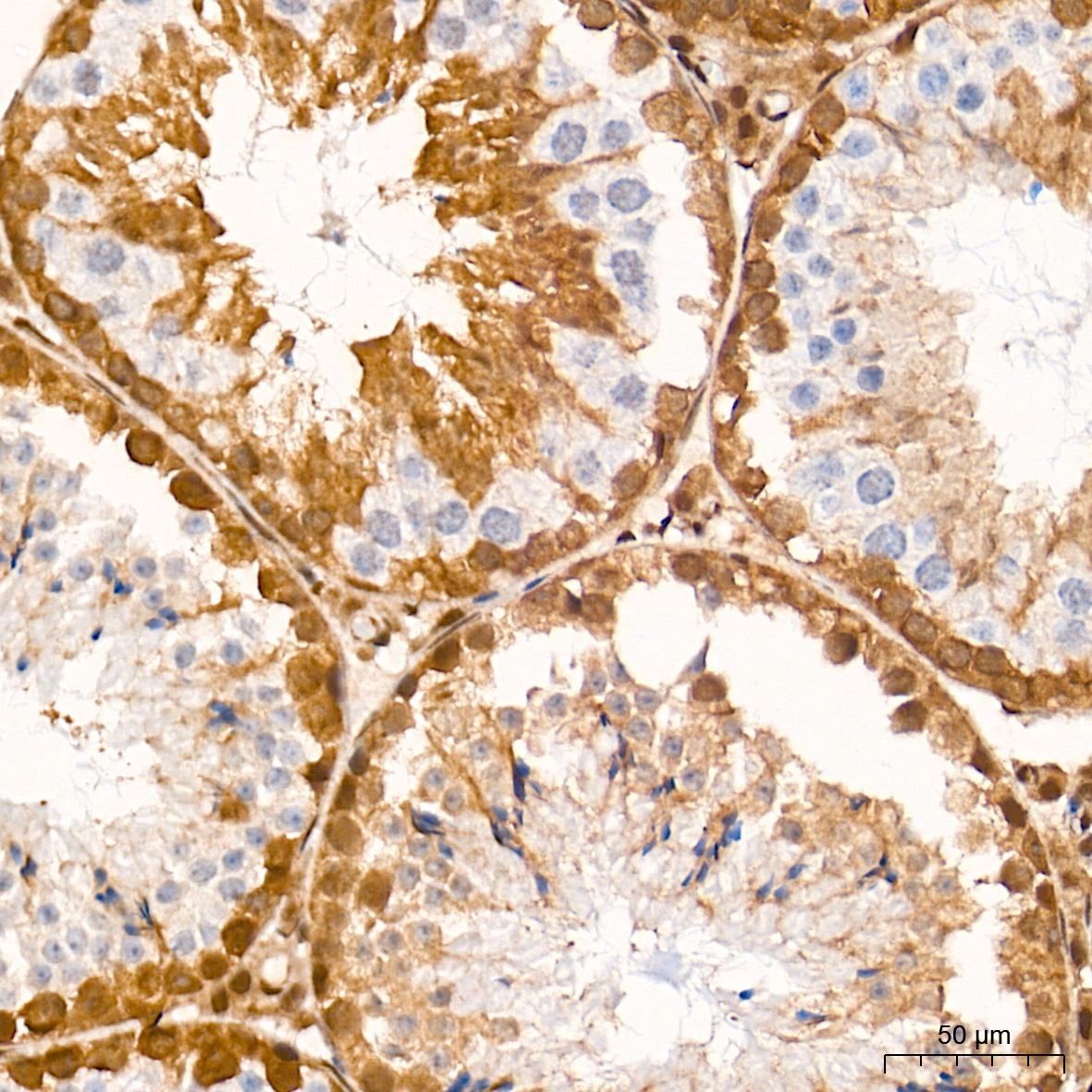
Immunohistochemistry analysis of paraffin-embedded Mouse testis tissue using [KO Validated] Smad4 Rabbit mAb (CAB19116) at a dilution of 1:500 (40x lens). High pressure antigen retrieval performed with 0.01M Tris-EDTA Buffer (pH 9.0) prior to IHC staining.
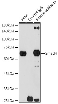
Immunoprecipitation analysis of 300 μg extracts of 293T cells using 3 μg Smad4 antibody (CAB19116). Western blot was performed from the immunoprecipitate using Smad4 antibody (CAB19116) at a dilution of1:1000.
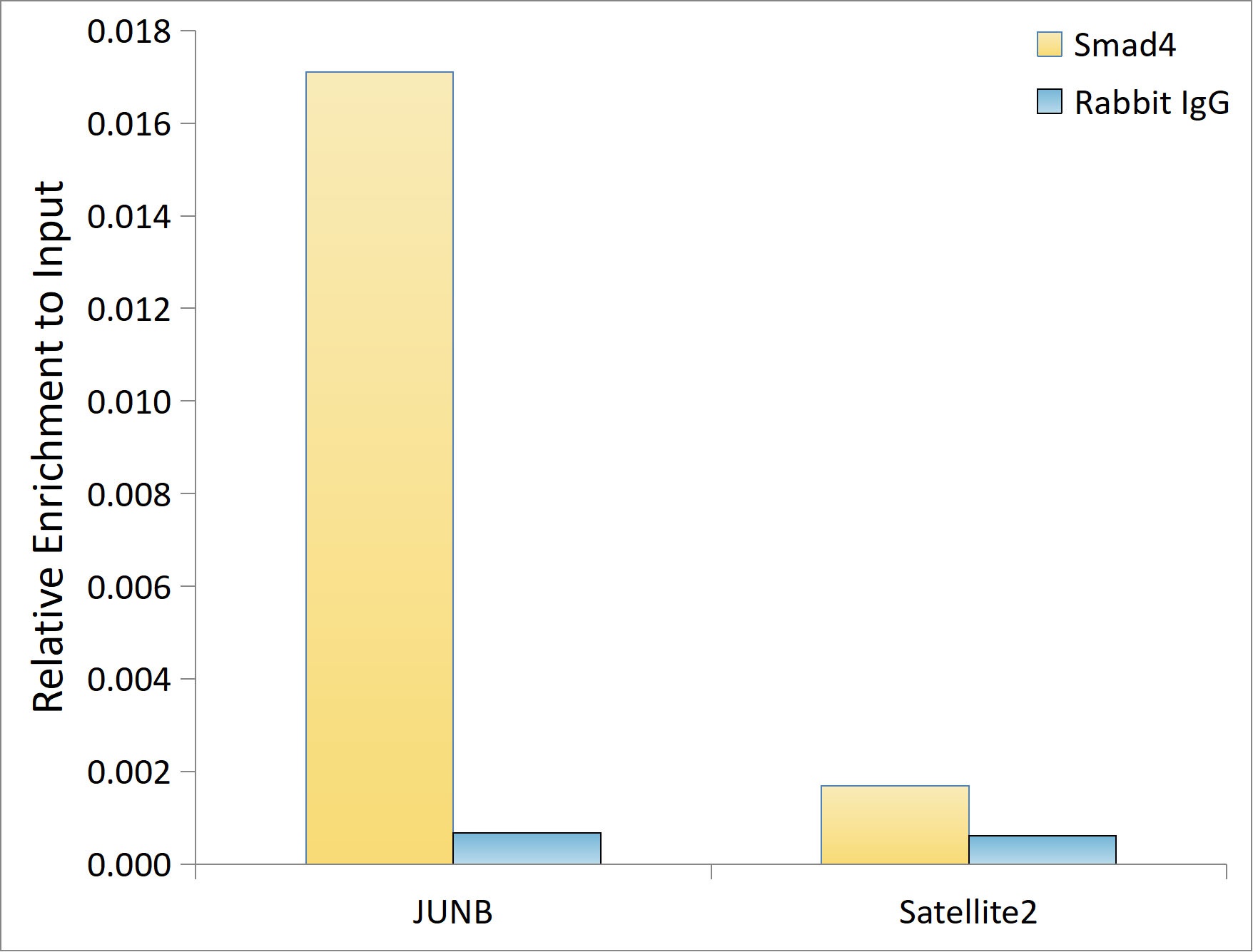
Chromatin immunoprecipitation analysis of extracts of HepG2 cells, using [KO Validated] Smad4 Rabbit mAb (CAB19116) and rabbit IgG.The amount of immunoprecipitated DNA was checked by quantitative PCR. Histogram was constructed by the ratios of the immunoprecipitated DNA to the input.
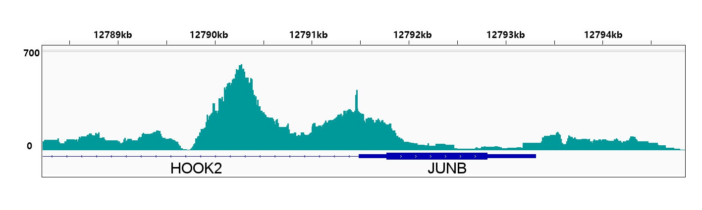
CUT&Tag was performed using the CUT&Tag Assay Kit (pAG-Tn5) for Illumina (RK20265) from10⁵ K562 with 1 μg of [KO Validated] Smad4 Rabbit mAb (CAB19116), followed by incubation with Goat Anti-Rabbit IgG(H+L)(AS070).The results denote the enrichment pattern of Smad4 around JUNB gene.