The KRR1 Antibody (CAB4487) is a high-quality antibody developed for reliable detection and analysis of target proteins. This antibody, generated in rabbits, is highly specific for human samples and has been validated for use in Western blot applications. By binding to the KRR1 protein, this antibody enables precise detection and analysis in a variety of cell types, making it indispensable for studies in molecular biology and genetics.KRR1 is essential for ribosome assembly and maturation, playing a crucial role in maintaining cellular functions that rely on proper protein synthesis.
This antibody is validated for use in WB, IF/ICC, ELISA applications and has demonstrated reactivity against Human, Mouse, Rat samples.
Product Name:
KRR1 Antibody
SKU:
CAB4487
Size:
20μL, 100μL
Reactivity:
Human, Mouse, Rat
Conjugate:
Unconjugated
Immunogen:
Recombinant protein (or fragment).This information is considered to be commercially sensitive.
Recommended starting concentration is 1 μg/mL. Please optimize the concentration based on your specific assay requirements.
Synonyms:
HRB2, RIP-1, KRR1
Positive Sample:
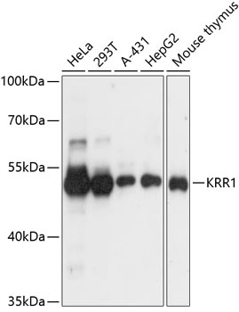
HeLa, 293T, A-431, HepG2, Mouse thymus
Cellular Localization:
Cytoplasm, Nucleus, Nucleolus.
Calculated MW:
44kDa
Observed MW:
44kDa
Enables RNA binding activity. Predicted to be involved in rRNA processing. Located in chromosome; intercellular bridge; and nuclear lumen.
Purification Method
Affinity purification
Gene ID
11103
RRID
AB_2765708
Buffer Information
Store at -20℃. Avoid freeze / thaw cycles. Buffer: PBS with 0.01% thimerosal,50% glycerol,pH7.3.
Western blot analysis of various lysates using KRR1 Rabbit pAb (CAB4487) at 1:3000 dilution. Secondary antibody: HRP-conjugated Goat anti-Rabbit IgG (H+L) (CABS014) at 1:10000 dilution. Lysates/proteins: 25μg per lane. Blocking buffer: 3% nonfat dry milk in TBST. Detection: ECL Basic Kit (AbGn00020). Exposure time: 1s.
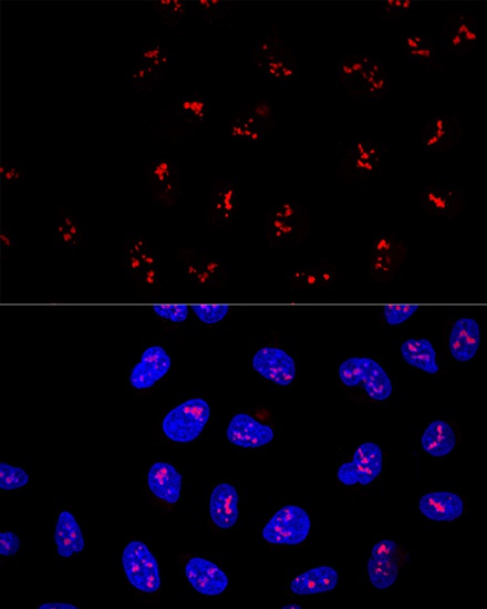
Confocal immunofluorescence analysis of U2OS cells using KRR1 Rabbit pAb (CAB4487) at dilution of 1:100. Blue: DAPI for nuclear staining.
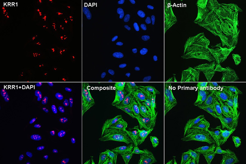
Confocal Immunofluorescent analysis of U2OS cells using KRR1 Rabbit pAb (CAB4487) at dilution of 1:100 (40x lens)(red). β-Actin (AC004: ACTB Mouse mAb) for Cytoskeleton staining(green), DAPI for nuclear staining(blue).