Krt18 Antibody is a premium polyclonal that offers outstanding performance and reliability for demanding research applications. Rigorously validated for ELISA, WB, IHC, this antibody ensures consistent, reproducible results across multiple experimental platforms. Demonstrates excellent reactivity with Mouse, Human samples, providing researchers with confidence in cross-species compatibility. Conveniently packaged in 50ug format to meet your experimental needs. For optimal performance, store at -20°C or -80°C and maintains stability for 12 months. Backed by rigorous quality control testing to ensure superior performance in your critical research applications.
Product Name:
Krt18 Antibody (PACO60745)
SKU:
PACO60745
Size:
50μg
Isotype:
IgG
Host Species:
Rabbit
Reactivity:
Mouse, Human
Immunogen:
Recombinant Mouse Keratin, type I cytoskeletal 18 protein (200-423AA)
Immunogen Species:
Mus musculus (Mouse)
Uniprot No:
P05784
Form:
Liquid
Tested Applications:
ELISAWBIHC
Recommended Dilution:
WB 1:500-1:5000, IHC 1:20-1:200
Synonyms:
Krt18 antibody, Kerd antibody, Krt1-18 antibody, Keratin antibody, type I cytoskeletal 18 antibody, Cytokeratin endo B antibody, Keratin D antibody, Cytokeratin-18 antibody, CK-18 antibody, Keratin-18 antibody, K18 antibody
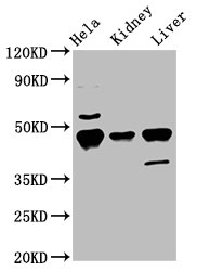
Western Blot Positive WB detected in: Hela whole cell lysate, Mouse kidney tissue, Mouse liver tissue All lanes: Krt18 antibody at 5.5µg/ml Secondary Goat polyclonal to rabbit IgG at 1/50000 dilution Predicted band size: 48 kDa Observed band size: 48 kDa
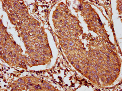
IHC image of PACO60745 diluted at 1:100 and staining in paraffin-embedded human cervical cancer performed on a Leica BondTM system. After dewaxing and hydration, antigen retrieval was mediated by high pressure in a citrate buffer (pH 6.0). Section was blocked with 10% normal goat serum 30min at RT. Then primary antibody (1% BSA) was incubated at 4°C overnight. The primary is detected by a biotinylated secondary antibody and visualized using an HRP conjugated SP system.
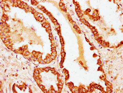
IHC image of PACO60745 diluted at 1:100 and staining in paraffin-embedded human prostate cancer performed on a Leica BondTM system. After dewaxing and hydration, antigen retrieval was mediated by high pressure in a citrate buffer (pH 6.0). Section was blocked with 10% normal goat serum 30min at RT. Then primary antibody (1% BSA) was incubated at 4°C overnight. The primary is detected by a biotinylated secondary antibody and visualized using an HRP conjugated SP system.


-S33 antibody . Secondary antibody: HRP Goat Anti-Rabbit IgG (H+L) at 1:10000 dilution. Lysates/proteins: 25ug per lane. Blocking buffer: 3% BSA.")

