The LASP1 Antibody (CAB3941) is a high-quality antibody developed for reliable detection and analysis of target proteins. This antibody, generated in rabbits, shows high specificity and reactivity towards LASP1 in human samples, making it ideal for Western blotting applications. By targeting LASP1, researchers can investigate its role in cancer metastasis, cell motility, and cytoskeletal dynamics.LASP1, also known as LIM and SH3 domain protein 1, is a key player in cellular processes that drive tumor progression and invasion.
This antibody is validated for use in WB, IF/ICC, IP, ELISA applications and has demonstrated reactivity against Human, Mouse, Rat samples.
Product Name:
LASP1 Antibody
SKU:
CAB3941
Size:
20μL, 100μL
Reactivity:
Human, Mouse, Rat
Conjugate:
Unconjugated
Immunogen:
Recombinant protein (or fragment).This information is considered to be commercially sensitive.
This gene encodes a member of a subfamily of LIM proteins, characterized by a LIM motif and a domain of Src homology region 3, and also a member of the nebulin family of actin-binding proteins. The encoded protein is a cAMP and cGMP dependent signaling protein and binds to the actin cytoskeleton at extensions of the cell membrane. The encoded protein has been linked to metastatic breast cancer, hematopoetic tumors such as B-cell lymphomas, and colorectal cancer.
Purification Method
Affinity purification
Gene ID
3927
RRID
AB_2765401
Buffer Information
Store at -20℃. Avoid freeze / thaw cycles. Buffer: PBS containing 50% glycerol, preserved with proclin300 or sodium azide, pH 7.3.
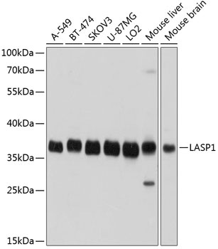
Western blot analysis of various lysates using LASP1 Rabbit pAb (CAB3941) at 1:1000 dilution. Secondary antibody: HRP-conjugated Goat anti-Rabbit IgG (H+L) (CABS014) at 1:10000 dilution. Lysates/proteins: 25μg per lane. Blocking buffer: 3% nonfat dry milk in TBST. Detection: ECL Basic Kit (AbGn00020). Exposure time: 1s.
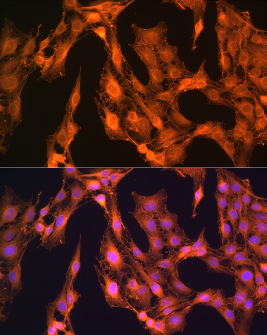
Immunofluorescence analysis of C6 cells using LASP1 Rabbit pAb (CAB3941) at dilution of 1:100 (40x lens). Secondary antibody: Cy3-conjugated Goat anti-Rabbit IgG (H+L) (CABS007) at 1:500 dilution. Blue: DAPI for nuclear staining.
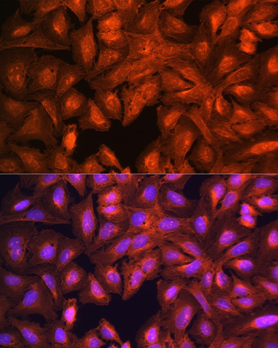
Immunofluorescence analysis of U-2 OS cells using LASP1 Rabbit pAb (CAB3941) at dilution of 1:100 (40x lens). Secondary antibody: Cy3-conjugated Goat anti-Rabbit IgG (H+L) (CABS007) at 1:500 dilution. Blue: DAPI for nuclear staining.
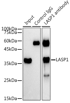
Immunoprecipitation analysis of 300 μg extracts of A-549 cells using 3 μg LASP1 antibody (CAB3941). Western blot was performed from the immunoprecipitate using LASP1 antibody (CAB3941) at a dilution of 1:1000.