The Galectin 3/LGALS3 Monoclonal Antibody (CAB11198) is a high-quality antibody developed for reliable detection and analysis of target proteins. This antibody, produced in rabbits, exhibits high reactivity with Galectin-3 in human samples and has been validated for use in Western blot applications. It specifically targets Galectin-3, allowing for precise detection and analysis in a variety of cell types, making it an excellent choice for studies in immunology, cancer research, and other fields.Galectin-3 is known for its roles in cell adhesion, proliferation, apoptosis, and inflammation, and dysregulation of its expression has been implicated in various diseases, including cancer, fibrosis, and cardiovascular disorders.
This antibody is validated for use in WB, IHC-P, IP, ELISA applications and has demonstrated reactivity against Human, Mouse, Rat samples.
Product Name:
Galectin 3/LGALS3 Monoclonal Antibody
SKU:
CAB11198
Size:
20μL, 100μL
Reactivity:
Human, Mouse, Rat
Clone Number:
ARC0542
Conjugate:
Unconjugated
Immunogen:
Synthetic peptide. This information is considered to be commercially sensitive.
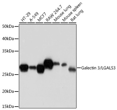
HT-29, A549, MCF7, RAW 264.7, Mouse lung, Mouse spleen, Rat lung
Cellular Localization:
Cytoplasm, Nucleus, Secreted.
Calculated MW:
26kDa
Observed MW:
28kDa
This gene encodes a member of the galectin family of carbohydrate binding proteins. Members of this protein family have an affinity for beta-galactosides. The encoded protein is characterized by an N-terminal proline-rich tandem repeat domain and a single C-terminal carbohydrate recognition domain. This protein can self-associate through the N-terminal domain allowing it to bind to multivalent saccharide ligands. This protein localizes to the extracellular matrix,the cytoplasm and the nucleus. This protein plays a role in numerous cellular functions including apoptosis,innate immunity,cell adhesion and T-cell regulation. The protein exhibits antimicrobial activity against bacteria and fungi. Alternate splicing results in multiple transcript variants.
Purification Method
Affinity purification
Gene ID
3958
RRID
AB_2861518
Buffer Information
Store at -20℃. Avoid freeze / thaw cycles. Buffer: PBS containing 50% glycerol and 0.05% BSA, preserved with proclin300 or sodium azide, pH 7.3.
Western blot analysis of various lysates using Galectin 3/LGALS3 Rabbit mAb (CAB11198) at 1:1000 dilution. Secondary antibody: HRP-conjugated Goat anti-Rabbit IgG (H+L) (CABS014) at 1:10000 dilution. Lysates/proteins: 25μg per lane. Blocking buffer: 3% nonfat dry milk in TBST. Detection: ECL Basic Kit (AbGn00020). Exposure time: 10s.
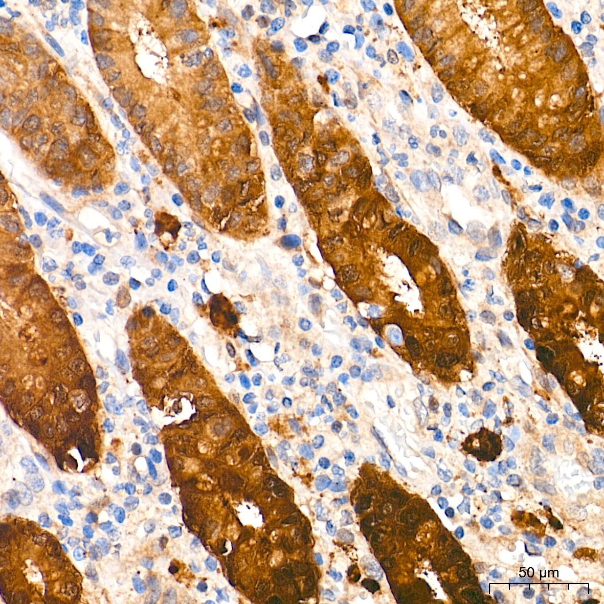
Immunohistochemistry analysis of paraffin-embedded Human colon tissue using Galectin 3/LGALS3 Rabbit mAb (CAB11198) at a dilution of 1:500 (40x lens). High pressure antigen retrieval performed with 0.01M Tris-EDTA Buffer (pH 9.0) prior to IHC staining.
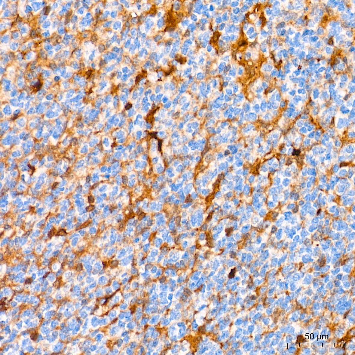
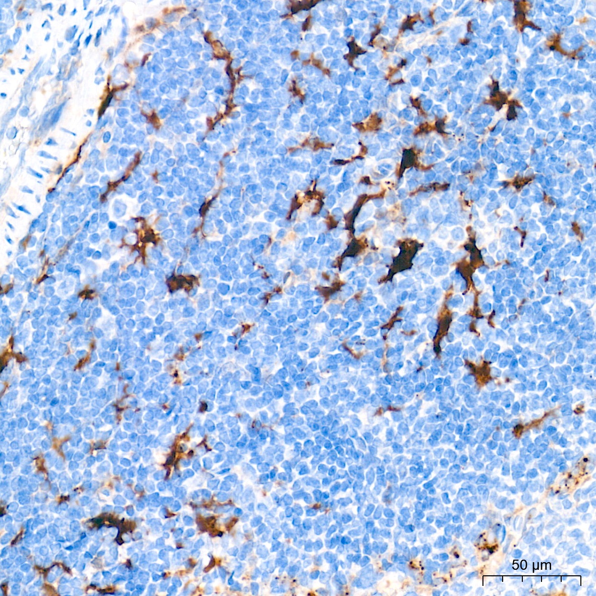
Immunohistochemistry analysis of paraffin-embedded Human tonsil tissue using Galectin 3/LGALS3 Rabbit mAb (CAB11198) at a dilution of 1:500 (40x lens). High pressure antigen retrieval performed with 0.01M Tris-EDTA Buffer (pH 9.0) prior to IHC staining.
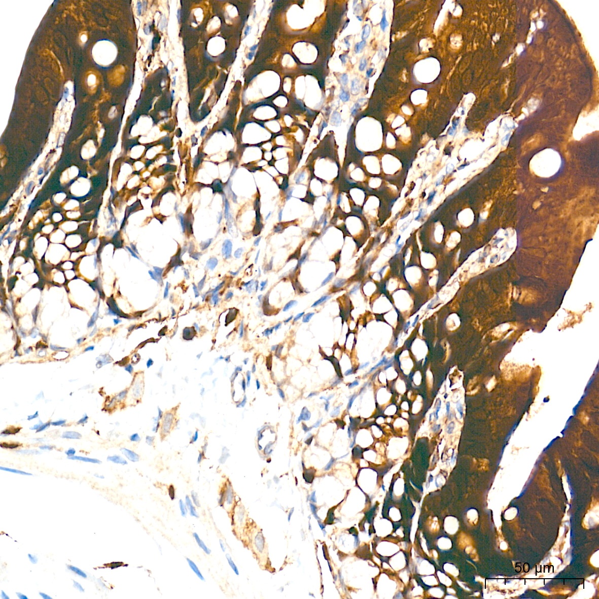
Immunohistochemistry analysis of paraffin-embedded Rat colon tissue using Galectin 3/LGALS3 Rabbit mAb (CAB11198) at a dilution of 1:500 (40x lens). High pressure antigen retrieval performed with 0.01M Tris-EDTA Buffer (pH 9.0) prior to IHC staining.
Immunohistochemistry analysis of paraffin-embedded Rat spleen tissue using Galectin 3/LGALS3 Rabbit mAb (CAB11198) at a dilution of 1:500 (40x lens). High pressure antigen retrieval performed with 0.01M Tris-EDTA Buffer (pH 9.0) prior to IHC staining.
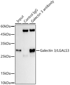
Immunoprecipitation analysis of 300 μg extracts of HT-29 cells using 3 μg Galectin 3/LGALS3 antibody (CAB11198). Western blot was performed from the immunoprecipitate using Galectin 3/LGALS3 antibody (CAB11198) at a dilution of 1:1000.