The LMBRD1 Antibody (CAB15866) is a high-quality antibody developed for reliable detection and analysis of target proteins. This antibody, generated in rabbits, exhibits high specificity and reactivity with human samples, making it ideal for use in Western blot applications. By targeting the LMBRD1 protein, this antibody allows for precise detection and analysis in a variety of cell types, making it essential for studies in the fields of biochemistry and molecular biology.
This antibody is validated for use in WB, IF/ICC, ELISA applications and has demonstrated reactivity against Human, Mouse samples.
Product Name:
LMBRD1 Antibody
SKU:
CAB15866
Size:
20μL, 100μL
Reactivity:
Human, Mouse
Conjugate:
Unconjugated
Immunogen:
Recombinant protein (or fragment).This information is considered to be commercially sensitive.
This gene encodes a lysosomal membrane protein that may be involved in the transport and metabolism of cobalamin. This protein also interacts with the large form of the hepatitis delta antigen and may be required for the nucleocytoplasmic shuttling of the hepatitis delta virus. Mutations in this gene are associated with the vitamin B12 metabolism disorder termed, homocystinuria-megaloblastic anemia complementation type F.
Purification Method
Affinity purification
Gene ID
55788
RRID
AB_2763294
Buffer Information
Store at -20℃. Avoid freeze / thaw cycles. Buffer: PBS with 0.01% thimerosal,50% glycerol,pH7.3.
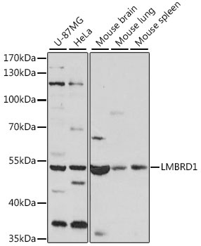
Western blot analysis of various lysates using LMBRD1 Rabbit pAb (CAB15866) at 1:1000 dilution. Secondary antibody: HRP-conjugated Goat anti-Rabbit IgG (H+L) (CABS014) at 1:10000 dilution. Lysates/proteins: 25μg per lane. Blocking buffer: 3% nonfat dry milk in TBST. Detection: ECL Basic Kit (AbGn00020). Exposure time: 5s.
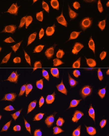
Immunofluorescence analysis of L929 cells using LMBRD1 Rabbit pAb (CAB15866) at dilution of 1:100. Secondary antibody: Cy3-conjugated Goat anti-Rabbit IgG (H+L) (CABS007) at 1:500 dilution. Blue: DAPI for nuclear staining.