The LMO2 Antibody (CAB1903) is a high-quality antibody developed for reliable detection and analysis of target proteins. This antibody, raised in rabbits, is highly specific to human Rhombotin 2 and is validated for use in immunostaining and Western blot applications. It binds specifically to Rhombotin 2, allowing for accurate detection and analysis in a variety of cell types.Rhombotin 2 is known for its involvement in the development and differentiation of blood cells, making it a crucial factor in understanding diseases such as leukemia and lymphoma.
This antibody is validated for use in WB, ELISA applications and has demonstrated reactivity against Human, Mouse, Rat samples.
Product Name:
LMO2 Antibody
SKU:
CAB1903
Size:
20μL, 100μL
Reactivity:
Human, Mouse, Rat
Conjugate:
Unconjugated
Immunogen:
Recombinant protein (or fragment).This information is considered to be commercially sensitive.
Recommended starting concentration is 1 μg/mL. Please optimize the concentration based on your specific assay requirements.
Synonyms:
TTG2, LMO-2, RBTN2, RHOM2, RBTNL1, LMO2
Positive Sample:
LO2, A-549, 293T, Mouse liver, Mouse kidney, Rat liver
Cellular Localization:
Nucleus.
Calculated MW:
18kDa
Observed MW:
18kDa
LMO2 encodes a cysteine-rich, two LIM-domain protein that is required for yolk sac erythropoiesis. The LMO2 protein has a central and crucial role in hematopoietic development and is highly conserved. The LMO2 transcription start site is located approximately 25 kb downstream from the 11p13 T-cell translocation cluster (11p13 ttc), where a number T-cell acute lymphoblastic leukemia-specific translocations occur. Alternative splicing results in multiple transcript variants encoding different isoforms.
Purification Method
Affinity purification
Gene ID
4005
RRID
AB_2763934
Buffer Information
Store at -20℃. Avoid freeze / thaw cycles. Buffer: PBS with 0.01% thimerosal,50% glycerol,pH7.3.
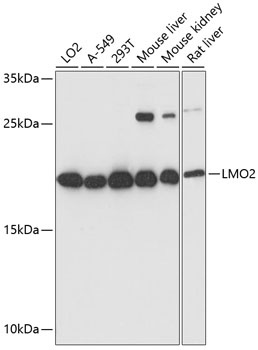
Western blot analysis of various lysates using LMO2 Rabbit pAb (CAB1903) at 1:3000 dilution. Secondary antibody: HRP-conjugated Goat anti-Rabbit IgG (H+L) (CABS014) at 1:10000 dilution. Lysates/proteins: 25μg per lane. Blocking buffer: 3% nonfat dry milk in TBST. Detection: ECL Basic Kit (AbGn00020). Exposure time: 30s.