The LNPEP Antibody (CAB11959) is a high-quality antibody developed for reliable detection and analysis of target proteins. This antibody, generated in rabbits, is highly specific for human samples and is validated for use in Western blot applications.LNPEP is known for its role in modulating immune responses by processing peptides involved in inflammation and immune cell activation. It has been implicated in various diseases, including autoimmune disorders and cancer, making it a promising target for therapeutic interventions.
This antibody is validated for use in WB, IHC-P, ELISA applications and has demonstrated reactivity against Human, Mouse, Rat samples.
Product Name:
LNPEP Antibody
SKU:
CAB11959
Size:
20μL, 100μL
Reactivity:
Human, Mouse, Rat
Conjugate:
Unconjugated
Immunogen:
Recombinant protein (or fragment).This information is considered to be commercially sensitive.
Recommended starting concentration is 1 μg/mL. Please optimize the concentration based on your specific assay requirements.
Synonyms:
CAP, IRAP, PLAP, P-LAP, LNPEP
Positive Sample:
Mouse brain,Rat brain
Cellular Localization:
Cell Membrane, Secreted, Single-Pass Type Ii Membrane Protein.
Calculated MW:
117kDa
Observed MW:
165kDa
This gene encodes a zinc-dependent aminopeptidase that cleaves vasopressin, oxytocin, lys-bradykinin, met-enkephalin, dynorphin A and other peptide hormones. The protein can be secreted in maternal serum, reside in intracellular vesicles with the insulin-responsive glucose transporter GLUT4, or form a type II integral membrane glycoprotein. The protein catalyzes the final step in the conversion of angiotensinogen to angiotensin IV (AT4) and is also a receptor for AT4. Alternative splicing results in multiple transcript variants encoding different isoforms.
Purification Method
Affinity purification
Gene ID
4012
RRID
AB_2758896
Buffer Information
Store at -20℃. Avoid freeze / thaw cycles. Buffer: PBS with 0.01% thimerosal,50% glycerol,pH7.3.
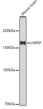
Western blot analysis of lysates from Mouse brain, using LNPEP Rabbit pAb (CAB11959) at 1:500 dilution. Secondary antibody: HRP-conjugated Goat anti-Rabbit IgG (H+L) (CABS014) at 1:10000 dilution. Lysates/proteins: 25μg per lane. Blocking buffer: 3% nonfat dry milk in TBST. Detection: ECL Basic Kit (AbGn00020). Exposure time: 180s.
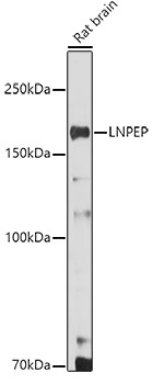
Western blot analysis of lysates from Rat brain, using LNPEP Rabbit pAb (CAB11959) at 1:500 dilution. Secondary antibody: HRP-conjugated Goat anti-Rabbit IgG (H+L) (CABS014) at 1:10000 dilution. Lysates/proteins: 25μg per lane. Blocking buffer: 3% nonfat dry milk in TBST. Detection: ECL Enhanced Kit (AbGn00021). Exposure time: 180s.
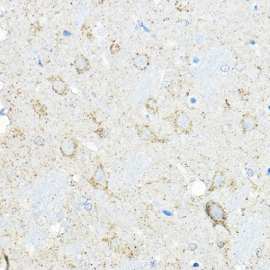
Immunohistochemistry analysis of paraffin-embedded Rat brain using LNPEP Rabbit pAb (CAB11959) at dilution of 1:100 (40x lens). High pressure antigen retrieval performed with 0.01M Citrate buffer (pH 6.0) prior to IHC staining.
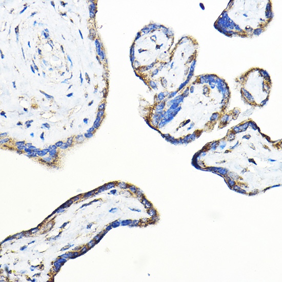
Immunohistochemistry analysis of paraffin-embedded Human placenta using LNPEP Rabbit pAb (CAB11959) at dilution of 1:100 (40x lens). High pressure antigen retrieval performed with 0.01M Citrate buffer (pH 6.0) prior to IHC staining.