The LRPPRC Antibody (CAB3365) is a high-quality antibody developed for reliable detection and analysis of target proteins. This antibody, produced in rabbits, is highly specific for human LRPPRC samples and has been extensively validated for Western blot applications.LRPPRC, also known as leucine rich PPR motif-containing protein, is essential for maintaining mitochondrial function and energy production in cells. Dysregulation of LRPPRC has been implicated in various diseases including metabolic disorders and cancer.
This antibody is validated for use in WB, IHC-P, IF/ICC, ELISA applications and has demonstrated reactivity against Human, Mouse, Rat samples.
Product Name:
LRPPRC Antibody
SKU:
CAB3365
Size:
20μL, 100μL
Reactivity:
Human, Mouse, Rat
Conjugate:
Unconjugated
Immunogen:
Recombinant protein (or fragment).This information is considered to be commercially sensitive.
This gene encodes a leucine-rich protein that has multiple pentatricopeptide repeats (PPR). The precise role of this protein is unknown but studies suggest it may play a role in cytoskeletal organization, vesicular transport, or in transcriptional regulation of both nuclear and mitochondrial genes. The protein localizes primarily to mitochondria and is predicted to have an N-terminal mitochondrial targeting sequence. Mutations in this gene are associated with the French-Canadian type of Leigh syndrome.
Purification Method
Affinity purification
Gene ID
10128
RRID
AB_2765075
Buffer Information
Store at -20℃. Avoid freeze / thaw cycles. Buffer: PBS containing 50% glycerol, preserved with proclin300 or sodium azide, pH 7.3.
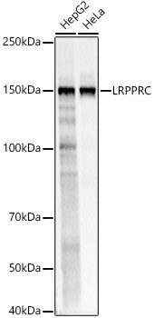
Western blot analysis of various lysates, using LRPPRC Rabbit pAb (CAB3365) at 1:2000 dilution. Secondary antibody: HRP-conjugated Goat anti-Rabbit IgG (H+L) (CABS014) at 1:10000 dilution. Lysates/proteins: 25μg per lane. Blocking buffer: 3% nonfat dry milk in TBST. Detection: ECL Basic Kit (AbGn00020). Exposure time: 0.5s.
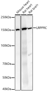
Western blot analysis of various lysates, using LRPPRC Rabbit pAb (CAB3365) at 1:2000 dilution. Secondary antibody: HRP-conjugated Goat anti-Rabbit IgG (H+L) (CABS014) at 1:10000 dilution. Lysates/proteins: 25μg per lane. Blocking buffer: 3% nonfat dry milk in TBST. Detection: ECL Basic Kit (AbGn00020). Exposure time: 10s.
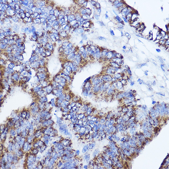
Immunohistochemistry analysis of paraffin-embedded Human colon carcinoma using LRPPRC Rabbit pAb (CAB3365) at dilution of 1:200 (40x lens). Microwave antigen retrieval performed with 0.01M PBS Buffer (pH 7.2) prior to IHC staining.
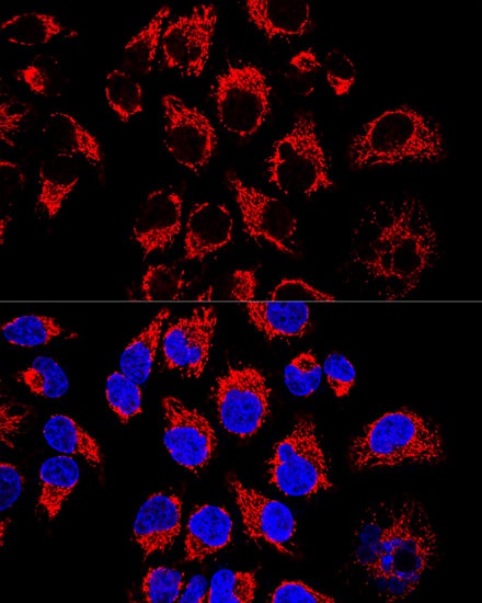
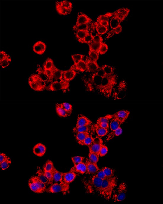
Confocal immunofluorescence analysis of Hela cells using LRPPRC Rabbit pAb (CAB3365) at dilution of 1:400. Blue: DAPI for nuclear staining.
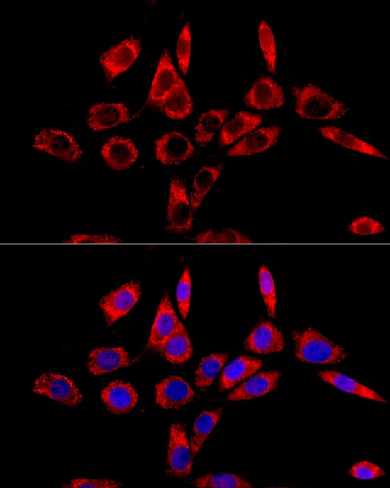
Immunofluorescence analysis of HepG2 cells using LRPPRC Rabbit pAb (CAB3365) at dilution of 1:100 (40x lens). Secondary antibody: Cy3-conjugated Goat anti-Rabbit IgG (H+L) (CABS007) at 1:500 dilution. Blue: DAPI for nuclear staining.
Immunofluorescence analysis of NIH/3T3 cells using LRPPRC Rabbit pAb (CAB3365) at dilution of 1:100 (40x lens). Secondary antibody: Cy3-conjugated Goat anti-Rabbit IgG (H+L) (CABS007) at 1:500 dilution. Blue: DAPI for nuclear staining.