The LSP1 Monoclonal Antibody (CAB3355) is a high-quality antibody developed for reliable detection and analysis of target proteins. This antibody, generated in rabbits, is highly specific and reactive with human samples, making it ideal for studying LSP1 in various experimental settings. LSP1, also known as lymphocyte-specific protein 1, plays a crucial role in regulating immune responses and cell migration in the immune system. By targeting LSP1, researchers can gain insights into its functions in inflammation, immune cell activation, and cancer progression.
This antibody is validated for use in WB, IF/ICC, ELISA, IF-P applications and has demonstrated reactivity against Human, Mouse, Rat samples.
Product Name:
LSP1 Monoclonal Antibody
SKU:
CAB3355
Size:
20μL, 100μL
Reactivity:
Human, Mouse, Rat
Clone Number:
ARC1952
Conjugate:
Unconjugated
Immunogen:
Synthetic peptide. This information is considered to be commercially sensitive.
This gene encodes an intracellular F-actin binding protein. The protein is expressed in lymphocytes, neutrophils, macrophages, and endothelium and may regulate neutrophil motility, adhesion to fibrinogen matrix proteins, and transendothelial migration. Alternative splicing results in multiple transcript variants encoding different isoforms.
Purification Method
Affinity purification
Gene ID
4046
RRID
AB_2863041
Buffer Information
Store at -20℃. Avoid freeze / thaw cycles. Buffer: PBS containing 50% glycerol and 0.05% BSA, preserved with proclin300 or sodium azide, pH 7.3.
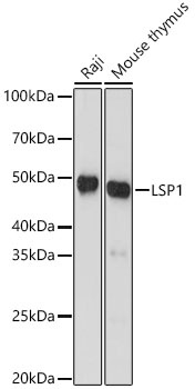
Western blot analysis of various lysates, using LSP1 Rabbit mAb (CAB3355) at 1:1000 dilution. Secondary antibody: HRP-conjugated Goat anti-Rabbit IgG (H+L) (CABS014) at 1:10000 dilution. Lysates/proteins: 25μg per lane. Blocking buffer: 3% nonfat dry milk in TBST. Detection: ECL Basic Kit (AbGn00020). Exposure time: 60s.
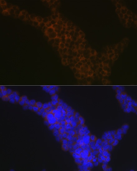
Immunofluorescence analysis of Jurkat cells using LSP1 Rabbit mAb (CAB3355) at dilution of 1:100 (40x lens). Secondary antibody: Cy3-conjugated Goat anti-Rabbit IgG (H+L) (CABS007) at 1:500 dilution. Blue: DAPI for nuclear staining.
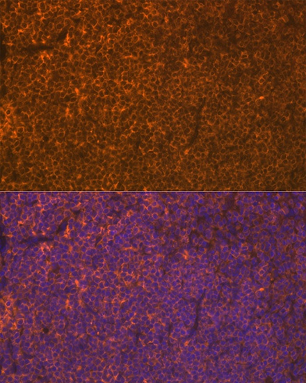
Immunofluorescence analysis of paraffin-embedded mouse spleen using LSP1 Rabbit mAb (CAB3355) at dilution of 1:100 (40x lens). Secondary antibody: Cy3-conjugated Goat anti-Rabbit IgG (H+L) (CABS007) at 1:500 dilution. Blue: DAPI for nuclear staining.