The LSP1 Antibody (CAB5617) is a high-quality antibody developed for reliable detection and analysis of target proteins. This antibody is raised in rabbits and is highly reactive with human samples, making it suitable for use in Western blot applications. By binding to the LSP1 protein, this antibody allows for the detection and analysis of LSP1 expression in various cell types.LSP1 is known for its role in mediating the interaction between immune cells and the extracellular matrix, as well as its involvement in cell adhesion and migration. Studies have shown that dysregulation of LSP1 expression can impact immune function and contribute to the development of inflammatory conditions and cancer.
This antibody is validated for use in WB, IHC-P, ELISA applications and has demonstrated reactivity against Human, Mouse samples.
Product Name:
LSP1 Antibody
SKU:
CAB5617
Size:
20μL, 100μL
Reactivity:
Human, Mouse
Conjugate:
Unconjugated
Immunogen:
Recombinant protein (or fragment).This information is considered to be commercially sensitive.
This gene encodes an intracellular F-actin binding protein. The protein is expressed in lymphocytes, neutrophils, macrophages, and endothelium and may regulate neutrophil motility, adhesion to fibrinogen matrix proteins, and transendothelial migration. Alternative splicing results in multiple transcript variants encoding different isoforms.
Purification Method
Affinity purification
Gene ID
4046
RRID
AB_2766378
Buffer Information
Store at -20℃. Avoid freeze / thaw cycles. Buffer: PBS containing 50% glycerol, preserved with proclin300 or sodium azide, pH 7.3.
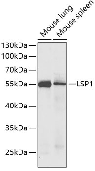
Western blot analysis of various lysates using LSP1 Rabbit pAb (CAB5617) at 1:1000 dilution. Secondary antibody: HRP-conjugated Goat anti-Rabbit IgG (H+L) (CABS014) at 1:10000 dilution. Lysates/proteins: 25μg per lane. Blocking buffer: 3% nonfat dry milk in TBST. Detection: ECL Enhanced Kit (AbGn00021). Exposure time: 30s.
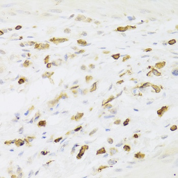
Immunohistochemistry analysis of paraffin-embedded Human gastric cancer using LSP1 Rabbit pAb (CAB5617) at dilution of 1:100 (40x lens). Microwave antigen retrieval performed with 0.01M PBS Buffer (pH 7.2) prior to IHC staining.