The LTA4H Monoclonal Antibody (CAB8918) is a high-quality antibody developed for reliable detection and analysis of target proteins. This antibody, generated in rabbits, exhibits high reactivity with human samples and has been validated for use in various applications such as Western blot and immunohistochemistry.LTA4H is a key player in the regulation of inflammation and immune responses, making it a promising target for studies in immunology and inflammation-related diseases. By binding to the LTA4H protein, this antibody enables the detection and analysis of LTA4H expression in different cell types, providing valuable insights for research into conditions such as asthma, rheumatoid arthritis, and inflammatory bowel disease.
This antibody is validated for use in WB, IF/ICC, ELISA applications and has demonstrated reactivity against Human, Mouse, Rat samples.
Product Name:
LTA4H Monoclonal Antibody
SKU:
CAB8918
Size:
20μL, 100μL
Reactivity:
Human, Mouse, Rat
Clone Number:
ARC1351
Conjugate:
Unconjugated
Immunogen:
Recombinant protein (or fragment).This information is considered to be commercially sensitive.
Recommended starting concentration is 1 μg/mL. Please optimize the concentration based on your specific assay requirements.
Synonyms:
LTA4H, leukotriene A4 hydrolase
Positive Sample:
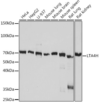
HeLa, Hep G2, U-937, Mouse lung, Mouse brain, Mouse spleen, Rat lung, Rat kidney
Cellular Localization:
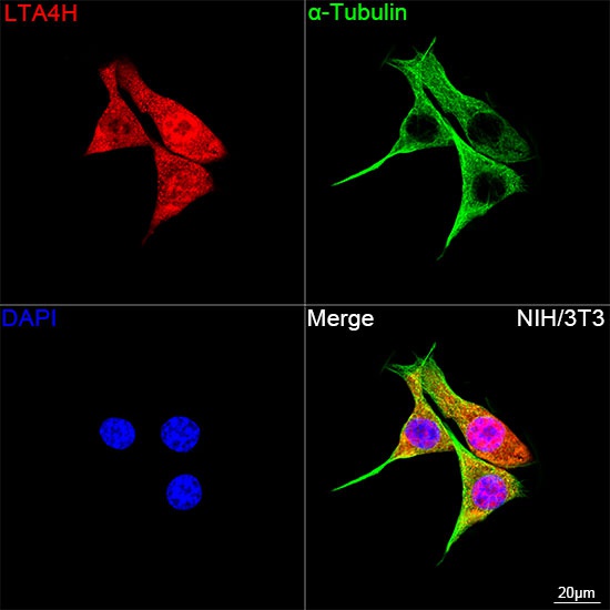
Cytoplasm.
Calculated MW:
69kDa
Observed MW:
70kDa
The protein encoded by this gene is an enzyme that contains both hydrolase and aminopeptidase activities. The hydrolase activity is used in the final step of the biosynthesis of leukotriene B4, a proinflammatory mediator. The aminopeptidase activity has been shown to degrade proline-glycine-proline (PGP), a neutrophil chemoattractant and biomarker for chronic obstructive pulmonary disease (COPD). Several transcript variants encoding different isoforms have been found for this gene.
Purification Method
Affinity purification
Gene ID
4048
RRID
AB_2863628
Buffer Information
Store at -20℃. Avoid freeze / thaw cycles. Buffer: PBS containing 50% glycerol and 0.05% BSA, preserved with proclin300 or sodium azide, pH 7.3.
Western blot analysis of various lysates using LTA4H Rabbit mAb (CAB8918) at 1:1000 dilution. Secondary antibody: HRP-conjugated Goat anti-Rabbit IgG (H+L) (CABS014) at 1:10000 dilution. Lysates/proteins: 25μg per lane. Blocking buffer: 3% nonfat dry milk in TBST. Detection: ECL Basic Kit (AbGn00020). Exposure time: 10s.
Confocal imaging of NIH/3T3 cells using LTA4H Rabbit mAb (CAB8918, dilution 1:200) followed by a further incubation with Cy3 Goat Anti-Rabbit IgG (H+L) (CABS007, dilution 1:500) (Red). The cells were counterstained with α-Tubulin Mouse mAb (AC012, dilution 1:400) followed by incubation with ABflo®488-conjugated Goat Anti-Mouse IgG (H+L) Ab (CABS076, dilution 1:500) (Green). DAPI was used for nuclear staining (Blue). Objective: 100x.