The LXRalpha Monoclonal Antibody (CAB3974) is a high-quality antibody developed for reliable detection and analysis of target proteins. This antibody, raised in rabbits, is highly specific for LXRalpha and has been validated for use in various applications, including immunohistochemistry and immunofluorescence.LXRalpha is a nuclear receptor that plays a crucial role in regulating cholesterol metabolism, inflammation, and immune response. Dysregulation of LXRalpha has been associated with various diseases, including atherosclerosis, diabetes, and inflammatory conditions.
This antibody is validated for use in WB, IHC-P, IF/ICC, ELISA applications and has demonstrated reactivity against Human, Mouse, Rat samples.
Product Name:
LXRalpha Monoclonal Antibody
SKU:
CAB3974
Size:
20μL, 100μL
Reactivity:
Human, Mouse, Rat
Clone Number:
ARC0877
Conjugate:
Unconjugated
Immunogen:
Synthetic peptide. This information is considered to be commercially sensitive.
Recommended starting concentration is 1 μg/mL. Please optimize the concentration based on your specific assay requirements.
Synonyms:
LXRA, LXR-a, RLD-1, LXRα
Positive Sample:
293T, HepG2, Mouse liver, Mouse kidney, Mouse stomach, Rat liver
Cellular Localization:
Nucleus.
Calculated MW:
50kDa
Observed MW:
50kDa
The protein encoded by this gene belongs to the NR1 subfamily of the nuclear receptor superfamily. The NR1 family members are key regulators of macrophage function, controlling transcriptional programs involved in lipid homeostasis and inflammation. This protein is highly expressed in visceral organs, including liver, kidney and intestine. It forms a heterodimer with retinoid X receptor (RXR), and regulates expression of target genes containing retinoid response elements. Studies in mice lacking this gene suggest that it may play an important role in the regulation of cholesterol homeostasis. Alternatively spliced transcript variants encoding different isoforms have been found for this gene.
Purification Method
Affinity purification
Gene ID
10062
RRID
AB_2863166
Buffer Information
Store at -20℃. Avoid freeze / thaw cycles. Buffer: PBS containing 50% glycerol and 0.05% BSA, preserved with proclin300 or sodium azide, pH 7.3.
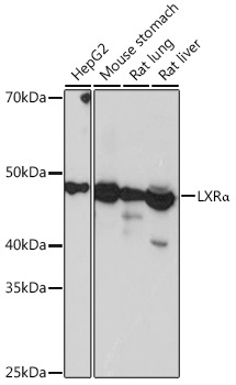
Western blot analysis of various lysates using LXRα Rabbit mAb (CAB3974) at 1:1000 dilution. Secondary antibody: HRP-conjugated Goat anti-Rabbit IgG (H+L) (CABS014) at 1:10000 dilution. Lysates/proteins: 25μg per lane. Blocking buffer: 3% nonfat dry milk in TBST. Detection: ECL Basic Kit (AbGn00020). Exposure time: 3s.
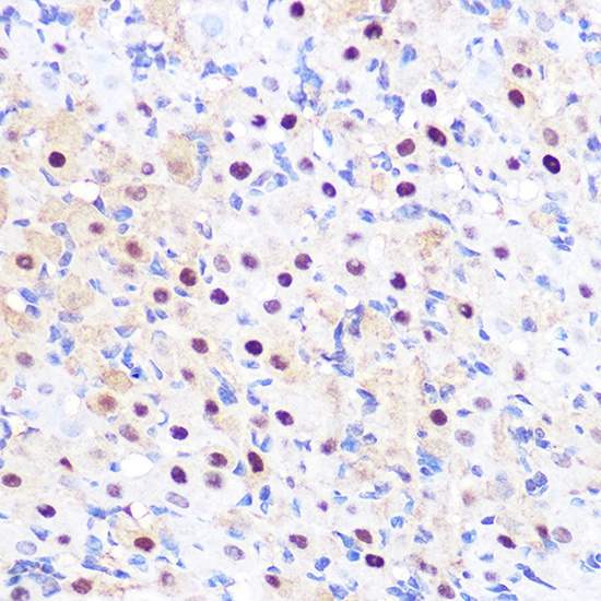
Immunohistochemistry analysis of paraffin-embedded Rat ovary using LXRα Rabbit mAb (CAB3974) at dilution of 1:100 (40x lens). Microwave antigen retrieval performed with 0.01M PBS Buffer (pH 7.2) prior to IHC staining.
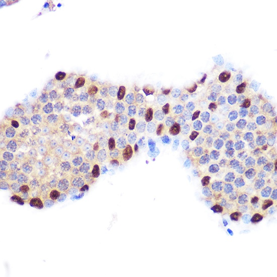
Immunohistochemistry analysis of paraffin-embedded Mouse testis using LXRα Rabbit mAb (CAB3974) at dilution of 1:100 (40x lens). Microwave antigen retrieval performed with 0.01M PBS Buffer (pH 7.2) prior to IHC staining.
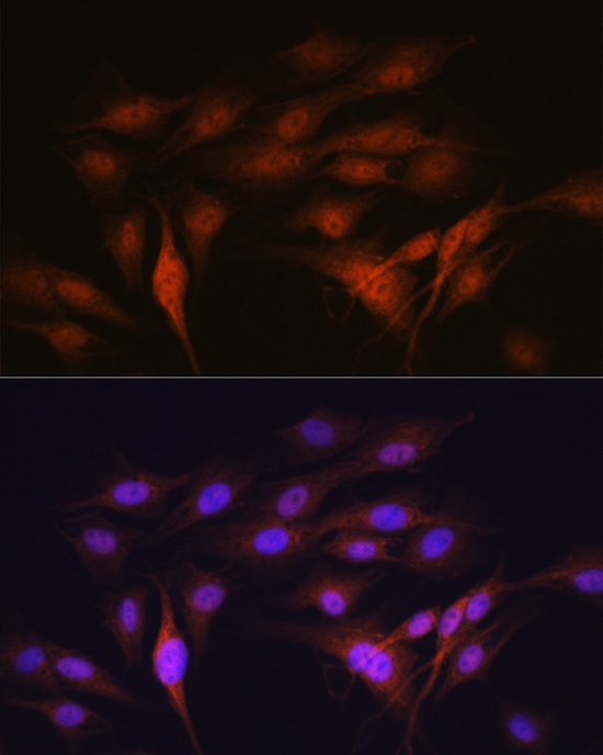
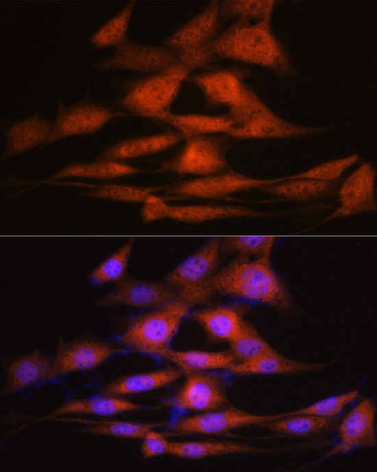
Immunofluorescence analysis of C6 cells using LXRα Rabbit mAb (CAB3974) at dilution of 1:100 (40x lens). Secondary antibody: Cy3-conjugated Goat anti-Rabbit IgG (H+L) (CABS007) at 1:500 dilution. Blue: DAPI for nuclear staining.
Immunofluorescence analysis of NIH-3T3 cells using LXRα Rabbit mAb (CAB3974) at dilution of 1:100 (40x lens). Secondary antibody: Cy3-conjugated Goat anti-Rabbit IgG (H+L) (CABS007) at 1:500 dilution. Blue: DAPI for nuclear staining.
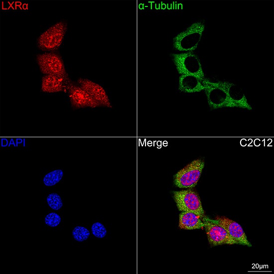
Confocal imaging of C2C12 cells using LXRα Rabbit mAb (CAB3974, dilution 1:100) followed by a further incubation with Cy3 Goat Anti-Rabbit IgG (H+L) (CABS007, dilution 1:500) (Red). The cells were counterstained with α-Tubulin Mouse mAb (AC012, dilution 1:400) followed by incubation with ABflo® 488-conjugated Goat Anti-Mouse IgG (H+L) Ab (CABS076, dilution 1:500) (Green). DAPI was used for nuclear staining (Blue). Objective: 100x.