M1-linkage Specific Polyubiquitin Antibody (CAB18200)
The M1-linkage Specific Polyubiquitin Antibody (CAB18200) is a high-quality antibody developed for reliable detection and analysis of target proteins. This antibody, raised in rabbits, is highly specific for M1-linked polyubiquitin chains and is validated for use in various applications, including Western blotting.Polyubiquitination, the process by which proteins are tagged with ubiquitin chains, plays a crucial role in protein degradation, signal transduction, and DNA repair. M1-linked polyubiquitin chains, in particular, are involved in immune signaling pathways and inflammatory responses, making them a key focus in immunology and cancer research.
This antibody is validated for use in WB, ELISA, DB applications and has demonstrated reactivity against Human, Mouse, Rat samples.
Product Name:
M1-linkage Specific Polyubiquitin Antibody
SKU:
CAB18200
Size:
20μL, 100μL
Reactivity:
Human, Mouse, Rat
Immunogen:
Synthetic peptide. This information is considered to be commercially sensitive.
Recommended starting concentration is 1 μg/mL. Please optimize the concentration based on your specific assay requirements.
Positive Sample:
HeLa, NIH/3T3, RAW264.7, C6, Mouse brain, Mouse kidney, Rat brain, Rat kidney
Observed MW:
Refertofigures
Ubiquitination,onetypeofthemostcommonpost-translationalmodification,mediatestheregulationofproteinhomeostasisinvivo. Substrate proteins can be modified with single ubiquitin moieties or with polymeric ubiquitin chains. Within polyubiquitin chains, ubiquitin can form eight different linkage types, using one of seven internal lysine residues (K6, K11, K27, K29, K33, K48, K63) or methionine at position 1 (M1).Here we focus on a distinct type of ubiquitination that is characterized by an inter-ubiquitin linkage through the N-terminal methionine, called M1-linked or linear ubiquitination. Formation, recognition, and disassembly of linear ubiquitin chains are highly specific processes that are implicated in immune signaling, cell death regulation and protein quality control. Consistent with their role in influencing signaling events, linear ubiquitin chains are formed in a transient and spatially regulated manner, making their detection and quantification challenging.
Purification Method
Affinity purification
RRID
AB_2861977
Buffer Information
Store at -20℃. Avoid freeze / thaw cycles. Buffer: PBS with 0.09% sodium azide,50% glycerol,pH7.3.
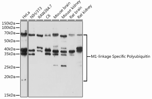
Western blot analysis of various lysates using M1-linkage Specific Polyubiquitin Rabbit pAb (CAB18200) at 1:1000 dilution. Secondary antibody: HRP-conjugated Goat anti-Rabbit IgG (H+L) (CABS014) at 1:10000 dilution. Lysates/proteins: 25μg per lane. Blocking buffer: 3% nonfat dry milk in TBST. Detection: ECL Basic Kit (AbGn00020). Exposure time: 90s.
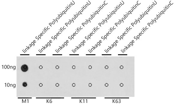
Dot-blot analysis of all sorts of peptides using M1-linkage Specific Polyubiquitin antibody (CAB18200) at 1:1000 dilution.