MAD2L1 binding protein Monoclonal Antibody (CAB19600)
The MAD2L1 binding protein Monoclonal Antibody (CAB19600) is a high-quality antibody developed for reliable detection and analysis of target proteins. This antibody, produced in rabbits, is highly specific for human samples and has been validated for use in Western blot applications. By binding to the MAD2L1BP protein, this antibody allows for precise detection and analysis in a variety of cell types, making it an essential component for research in molecular biology and cancer biology.MAD2L1BP, also known as MAD2L1 binding protein, is involved in the regulation of the cell cycle and ensures accurate distribution of chromosomes during cell division.
This antibody is validated for use in IHC-P, IF/ICC, ELISA applications and has demonstrated reactivity against Human, Mouse samples.
Product Name:
MAD2L1 binding protein Monoclonal Antibody
SKU:
CAB19600
Size:
20μL, 100μL
Reactivity:
Human, Mouse
Clone Number:
ARC2187
Conjugate:
Unconjugated
Immunogen:
A synthetic peptide corresponding to a sequence within amino acids 175-274 of human MAD2L1BP (Q15013).
The protein encoded by this gene was identified as a binding protein of the MAD2 mitotic arrest deficient-like 1 (MAD2/MAD2L1). MAD2 is a key component of the spindle checkpoint that delays the onset of anaphase until all the kinetochores are attached to the spindle. This protein may interact with the spindle checkpoint and coordinate cell cycle events in late mitosis. Alternatively spliced transcript variants encoding distinct isoforms have been observed.
Purification Method
Affinity purification
Gene ID
9587
Buffer Information
Store at -20℃. Avoid freeze / thaw cycles. Buffer: PBS containing 50% glycerol and 0.05% BSA, preserved with proclin300 or sodium azide, pH 7.3.
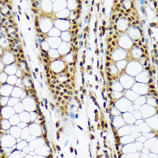
Immunohistochemistry analysis of paraffin-embedded Human colon using MAD2L1BP Rabbit mAb (CAB19600) at dilution of 1:100 (40x lens). Microwave antigen retrieval performed with 0.01M Tris/EDTA Buffer (pH 9.0) prior to IHC staining.
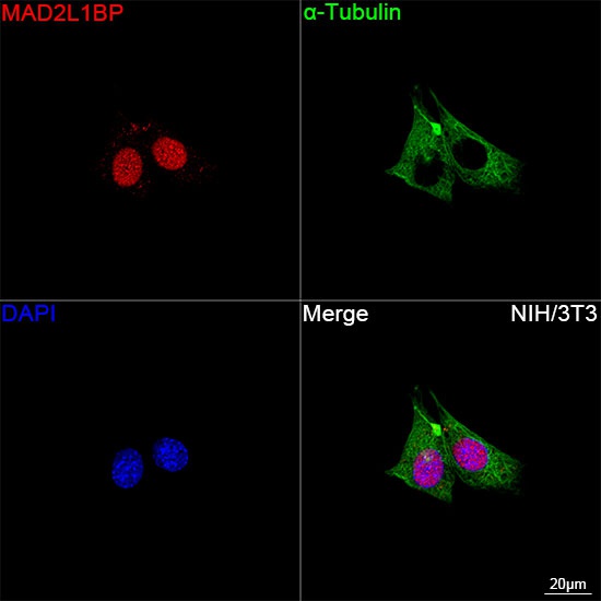
Confocal imaging of NIH/3T3 cells using MAD2L1BP Rabbit mAb (CAB19600, dilution 1:100) followed by a further incubation with Cy3 Goat Anti-Rabbit IgG (H+L) (CABS007, dilution 1:500) (Red). The cells were counterstained with α-Tubulin Mouse mAb (AC012, dilution 1:400) followed by incubation with ABflo® 488-conjugated Goat Anti-Mouse IgG (H+L) Ab (CABS076, dilution 1:500) (Green). DAPI was used for nuclear staining (Blue). Objective: 100x.


 / FOLR1 Rabbit Monoclonal Antibody (CAB22481)")


