The MAD2B/MAD2L2 Monoclonal Antibody (CAB4630) is a high-quality antibody developed for reliable detection and analysis of target proteins. This antibody specifically targets MAD2L2, a key protein involved in the regulation of cell cycle progression and genomic stability.Raised in rabbits, this monoclonal antibody is highly reactive with human samples and has been validated for use in various applications, including Western blotting and immunofluorescence. By binding to MAD2L2, researchers can accurately detect and analyze the protein in different cell types, providing valuable insights into its functions and regulatory mechanisms.MAD2L2 is known for its role in mitotic spindle assembly and the control of chromosome segregation, making it a crucial player in maintaining genomic integrity.
This antibody is validated for use in WB, IF/ICC, ELISA applications and has demonstrated reactivity against Human, Mouse, Rat samples.
Product Name:
MAD2B/MAD2L2 Monoclonal Antibody
SKU:
CAB4630
Size:
20μL, 100μL
Reactivity:
Human, Mouse, Rat
Clone Number:
ARC1126
Conjugate:
Unconjugated
Immunogen:
Synthetic peptide. This information is considered to be commercially sensitive.
Recommended starting concentration is 1 μg/mL. Please optimize the concentration based on your specific assay requirements.
Synonyms:
REV7, FANCV, MAD2B, POLZ2, MAD2B/MAD2L2
Positive Sample:
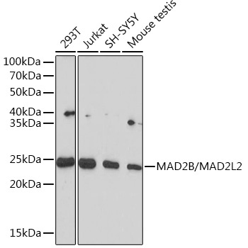
293T, Jurkat , SH-SY5Y, Mouse testis
Cellular Localization:
Cytoplasm, Nucleus, Cytoskeleton, Spindle.
Calculated MW:
24kDa
Observed MW:
24kDa
The protein encoded by this gene is a component of the mitotic spindle assembly checkpoint that prevents the onset of anaphase until all chromosomes are properly aligned at the metaphase plate. The encoded protein, which is similar to MAD2L1, is capable of interacting with ADAM9, ADAM15, REV1, and REV3 proteins.
Purification Method
Affinity purification
Gene ID
10459
RRID
AB_2863313
Buffer Information
Store at -20℃. Avoid freeze / thaw cycles. Buffer: PBS containing 50% glycerol and 0.05% BSA, preserved with proclin300 or sodium azide, pH 7.3.
Western blot analysis of various lysates using MAD2B/MAD2L2 Rabbit mAb (CAB4630) at 1:1000 dilution. Secondary antibody: HRP-conjugated Goat anti-Rabbit IgG (H+L) (CABS014) at 1:10000 dilution. Lysates/proteins: 25μg per lane. Blocking buffer: 3% nonfat dry milk in TBST. Detection: ECL Basic Kit (AbGn00020). Exposure time: 90s.
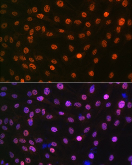
Immunofluorescence analysis of C6 cells using MAD2B/MAD2L2 Rabbit mAb (CAB4630) at dilution of 1:100 (40x lens). Secondary antibody: Cy3-conjugated Goat anti-Rabbit IgG (H+L) (CABS007) at 1:500 dilution. Blue: DAPI for nuclear staining.