The MAGEH1 Antibody (CAB17143) is a high-quality antibody developed for reliable detection and analysis of target proteins. This gene belongs to the non-CT (non cancer/testis) subgroup of the melanoma-associated antigen (MAGE) superfamily. The encoded protein is likely associated with apoptosis, cell cycle arrest, growth inhibition or cell differentiation. The protein may be involved in the atRA (all-trans retinoic acid) signaling through the STAT1-alpha (signal transducer and activator of transcription 1-alpha) pathway. RRID AB_2770250 Gene ID 28986 Swiss Prot Synonym APR1; APR-1; MAGEH; MAGEH1
This antibody is validated for use in WB, ELISA applications and has demonstrated reactivity against Mouse samples.
Product Name:
MAGEH1 Antibody
SKU:
CAB17143
Size:
100μL, 20μL
Reactivity:
Mouse
Clone Number:
-
Conjugate:
-
Immunogen:
Recombinant protein (or fragment).This information is considered to be commercially sensitive.
Tested Applications:
WBELISA
Recommended Dilution:
WB
1:500 - 1:2000
ELISA
Recommended starting concentration is 1 μg/mL. Please optimize the concentration based on your specific assay requirements.
Synonyms:
APR1, APR-1, MAGEH, MAGEH1
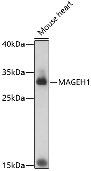
Positive Sample:
Mouse heart
Cellular Localization:
Nucleus.
Calculated MW:
24kDa
Observed MW:
28kDa
This gene belongs to the non-CT (non cancer/testis) subgroup of the melanoma-associated antigen (MAGE) superfamily. The encoded protein is likely associated with apoptosis, cell cycle arrest, growth inhibition or cell differentiation. The protein may be involved in the atRA (all-trans retinoic acid) signaling through the STAT1-alpha (signal transducer and activator of transcription 1-alpha) pathway. RRID AB_2770250 Gene ID 28986 Swiss Prot Synonym APR1; APR-1; MAGEH; MAGEH1
Purification Method:
Affinity purification
Gene ID:
28986
RRID:
AB_2770250
Buffer Information:
Store at -20℃. Avoid freeze / thaw cycles. Buffer: PBS with 0.01% thimerosal,50% glycerol,pH7.3.
Western blot analysis of lysates from mouse heart, using MAGEH1 Rabbit pAb (CAB17143) at 1:1000 dilution. Secondary antibody: HRP-conjugated Goat anti-Rabbit IgG (H+L) (AS014) at 1:10000 dilution. Lysates/proteins: 25μg per lane. Blocking buffer: 3% nonfat dry milk in TBST. Detection: ECL Basic Kit (AbGn00020). Exposure time: 180s.