The MAGOH Antibody (CAB6035) is a high-quality antibody developed for reliable detection and analysis of target proteins. This polyclonal antibody, generated in rabbits, exhibits high reactivity with human samples and has been validated for use in Western blot applications. By specifically binding to the MAGOH protein, this antibody enables accurate detection and analysis in a variety of cell types, making it an essential asset for studies in molecular biology and RNA biology research.MAGOH is a crucial component of the exon junction complex (EJC) and is known for its role in regulating mRNA translocation and translation. Its involvement in RNA processing makes it an intriguing target for investigations into gene expression, splicing efficiency, and cellular function.
This antibody is validated for use in WB, ELISA applications and has demonstrated reactivity against Human, Mouse, Rat samples.
Product Name:
MAGOH Antibody
SKU:
CAB6035
Size:
20μL, 100μL
Reactivity:
Human, Mouse, Rat
Conjugate:
Unconjugated
Immunogen:
Recombinant protein (or fragment).This information is considered to be commercially sensitive.
Recommended starting concentration is 1 μg/mL. Please optimize the concentration based on your specific assay requirements.
Synonyms:
MAGOH1, MAGOHA, MAGOH
Positive Sample:
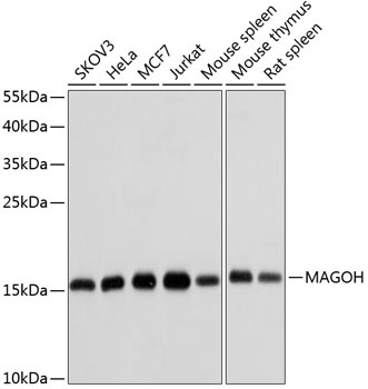
SKOV3, HeLa, MCF7, Jurkat, Mouse spleen, Mouse thymus, Rat spleen
Cellular Localization:
Cytoplasm, Nucleus, Nucleus Speckle.
Calculated MW:
17kDa
Observed MW:
17kDa
Drosophila that have mutations in their mago nashi (grandchildless) gene produce progeny with defects in germplasm assembly and germline development. This gene encodes the mammalian mago nashi homolog. In mammals, mRNA expression is not limited to the germ plasm, but is expressed ubiquitously in adult tissues and can be induced by serum stimulation of quiescent fibroblasts.
Purification Method
Affinity purification
Gene ID
4116
RRID
AB_2766723
Buffer Information
Store at -20℃. Avoid freeze / thaw cycles. Buffer: PBS containing 50% glycerol, preserved with proclin300 or sodium azide, pH 7.3.
Western blot analysis of various lysates using MAGOH Rabbit pAb (CAB6035) at 1:1000 dilution. Secondary antibody: HRP-conjugated Goat anti-Rabbit IgG (H+L) (CABS014) at 1:10000 dilution. Lysates/proteins: 25μg per lane. Blocking buffer: 3% nonfat dry milk in TBST. Detection: ECL Basic Kit (AbGn00020). Exposure time: 10s.