The MVP Antibody (CAB1980) is a high-quality antibody developed for reliable detection and analysis of target proteins. This antibody is produced in rabbits and shows high reactivity to human samples, making it suitable for Western blot applications. By binding specifically to MVP, this antibody enables accurate detection and analysis of MVP expression in a variety of cell types, making it an essential tool for studies in the fields of drug resistance, cellular transport, and cancer research.
This antibody is validated for use in WB, IHC-P, ELISA applications and has demonstrated reactivity against Human, Mouse samples.
Product Name:
MVP Antibody
SKU:
CAB1980
Size:
20μL, 100μL
Reactivity:
Human, Mouse
Conjugate:
Unconjugated
Immunogen:
Recombinant protein (or fragment).This information is considered to be commercially sensitive.
Recommended starting concentration is 1 μg/mL. Please optimize the concentration based on your specific assay requirements.
Synonyms:
LRP, VAULT1, MVP
Positive Sample:
NIH/3T3
Cellular Localization:
Cytoplasm, Nucleus, Nuclear Pore Complex.
Calculated MW:
99kDa
Observed MW:
110kDa
This gene encodes the major component of the vault complex. Vaults are multi-subunit ribonucleoprotein structures that may be involved in nucleo-cytoplasmic transport. The encoded protein may play a role in multiple cellular processes by regulating the MAP kinase, JAK/STAT and phosphoinositide 3-kinase/Akt signaling pathways. The encoded protein also plays a role in multidrug resistance, and expression of this gene may be a prognostic marker for several types of cancer. Alternatively spliced transcript variants have been observed for this gene.
Purification Method
Affinity purification
Gene ID
9961
RRID
AB_2764006
Buffer Information
Store at -20℃. Avoid freeze / thaw cycles. Buffer: PBS containing 50% glycerol, preserved with proclin300 or sodium azide, pH 7.3.
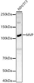
Western blot analysis of lysates from NIH/3T3 cells, using MVP Rabbit pAb (CAB1980) at 1:2000 dilution. Secondary antibody: HRP-conjugated Goat anti-Rabbit IgG (H+L) (CABS014) at 1:10000 dilution. Lysates/proteins: 25μg per lane. Blocking buffer: 3% nonfat dry milk in TBST. Detection: ECL Basic Kit (AbGn00020). Exposure time: 90s.
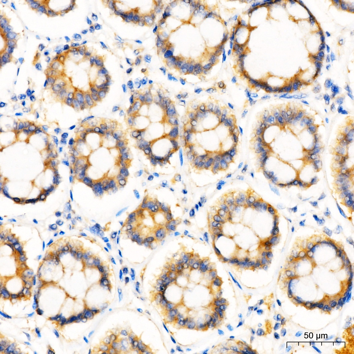
Immunohistochemistry analysis of paraffin-embedded Human colon tissue using MVP Rabbit pAb (CAB1980) at a dilution of 1:200 (40x lens). High pressure antigen retrieval was performed with 0.01 M Tris-EDTA buffer (pH 9.0) prior to IHC staining.