The Map2 Antibody (CAB0453) is a high-quality antibody developed for reliable detection and analysis of target proteins. MAP2 is a key structural protein found in neuronal cells, playing a crucial role in stabilizing microtubules and promoting neuronal development and plasticity.Raised in rabbits, this antibody offers high specificity and sensitivity when detecting MAP2 in human samples, making it suitable for use in Western blot applications. By binding to the MAP2 protein, researchers can effectively analyze and study its expression and function in different cell types, providing valuable insights into neurobiology and neurodegenerative diseases.
This antibody is validated for use in WB, IHC-P, ELISA, IF-P applications and has demonstrated reactivity against Mouse, Rat samples.
Product Name:
Map2 Antibody
SKU:
CAB0453
Size:
20μL, 100μL
Reactivity:
Mouse, Rat
Conjugate:
Unconjugated
Immunogen:
Recombinant protein (or fragment).This information is considered to be commercially sensitive.
Recommended starting concentration is 1 μg/mL. Please optimize the concentration based on your specific assay requirements.
Synonyms:
MAP-2, MAP2A, MAP2B, MAP2C, MAP2
Positive Sample:
Mouse brain
Cellular Localization:
Cytoplasm, Cytoskeleton.
Calculated MW:
200kDa
Observed MW:
220-280kd(MAP2A、MAP2B)
This gene encodes a protein that belongs to the microtubule-associated protein family. The proteins of this family are thought to be involved in microtubule assembly, which is an essential step in neurogenesis. The products of similar genes in rat and mouse are neuron-specific cytoskeletal proteins that are enriched in dentrites, implicating a role in determining and stabilizing dentritic shape during neuron development. A number of alternatively spliced variants encoding distinct isoforms have been described.
Purification Method
Affinity purification
Gene ID
4133
RRID
AB_2757199
Buffer Information
Store at -20℃. Avoid freeze / thaw cycles. Buffer: PBS containing 50% glycerol, preserved with proclin300 or sodium azide, pH 7.3.
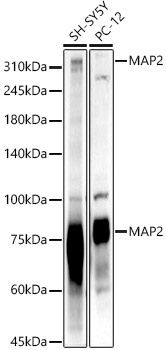
Western blot analysis of various lysates, using MAP2 Rabbit pAb (CAB0453) at 1:2000 dilution. Secondary antibody: HRP-conjugated Goat anti-Rabbit IgG (H+L) (CABS014) at 1:10000 dilution. Lysates/proteins: 25μg per lane. Blocking buffer: 3% nonfat dry milk in TBST. Detection: ECL Basic Kit (AbGn00020). Exposure time: 10s.
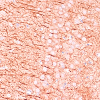
Immunohistochemistry analysis of paraffin-embedded Rat brain using MAP2 Rabbit pAb (CAB0453) at dilution of 1:100 (40x lens). Microwave antigen retrieval performed with 0.01M PBS Buffer (pH 7.2) prior to IHC staining.
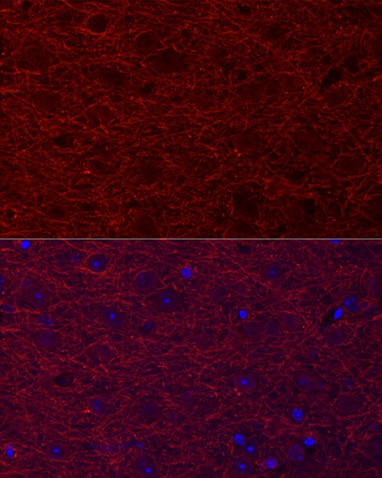
Immunofluorescence analysis of paraffin-embedded mouse brain using MAP2 Rabbit pAb (CAB0453) at dilution of 1:20 (40x lens). Secondary antibody: Cy3-conjugated Goat anti-Rabbit IgG (H+L) (CABS007) at 1:500 dilution. Blue: DAPI for nuclear staining.
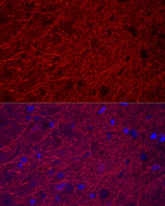
Immunofluorescence analysis of paraffin-embedded rat brain using MAP2 Rabbit pAb (CAB0453) at dilution of 1:20 (40x lens). Secondary antibody: Cy3-conjugated Goat anti-Rabbit IgG (H+L) (CABS007) at 1:500 dilution. Blue: DAPI for nuclear staining.