The MEK3 Antibody (CAB7718) is a high-quality antibody developed for reliable detection and analysis of target proteins. This antibody, generated in rabbits, exhibits high specificity and reactivity towards human samples, making it ideal for use in Western blot applications.MAP2K3, also known as dual specificity mitogen-activated protein kinase kinase 3, is involved in the MAPK signaling pathway and plays a crucial role in cell proliferation, differentiation, and apoptosis. Dysregulation of MAP2K3 has been linked to various diseases, including cancer, making it a potential target for therapeutic interventions.
This antibody is validated for use in WB, IHC-P, IF/ICC, ELISA applications and has demonstrated reactivity against Human, Mouse, Rat samples.
Product Name:
MEK3 Antibody
SKU:
CAB7718
Size:
20μL, 100μL
Reactivity:
Human, Mouse, Rat
Conjugate:
Unconjugated
Immunogen:
Recombinant protein (or fragment).This information is considered to be commercially sensitive.
Recommended starting concentration is 1 μg/mL. Please optimize the concentration based on your specific assay requirements.
Synonyms:
MEK3, MKK3, MAPKK3, PRKMK3, SAPKK2, SAPKK-2
Positive Sample:
Mouse heart, Mouse skeletal muscle
Cellular Localization:
Cytosol, Nucleoplasm.
Calculated MW:
39kDa
Observed MW:
39kDa
The protein encoded by this gene is a dual specificity protein kinase that belongs to the MAP kinase kinase family. This kinase is activated by mitogenic and environmental stress, and participates in the MAP kinase-mediated signaling cascade. It phosphorylates and thus activates MAPK14/p38-MAPK. This kinase can be activated by insulin, and is necessary for the expression of glucose transporter. Expression of RAS oncogene is found to result in the accumulation of the active form of this kinase, which thus leads to the constitutive activation of MAPK14, and confers oncogenic transformation of primary cells. The inhibition of this kinase is involved in the pathogenesis of Yersina pseudotuberculosis. Multiple alternatively spliced transcript variants that encode distinct isoforms have been reported for this gene.
Purification Method
Affinity purification
Gene ID
5606
RRID
AB_2770263
Buffer Information
Store at -20℃. Avoid freeze / thaw cycles. Buffer: PBS with 0.01% thimerosal,50% glycerol,pH7.3.
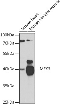
Western blot analysis of various lysates using MEK3 Rabbit pAb (CAB7718) at 1:1000 dilution. Secondary antibody: HRP-conjugated Goat anti-Rabbit IgG (H+L) (CABS014) at 1:10000 dilution. Lysates/proteins: 25μg per lane. Blocking buffer: 3% nonfat dry milk in TBST. Detection: ECL Basic Kit (AbGn00020). Exposure time: 90s.
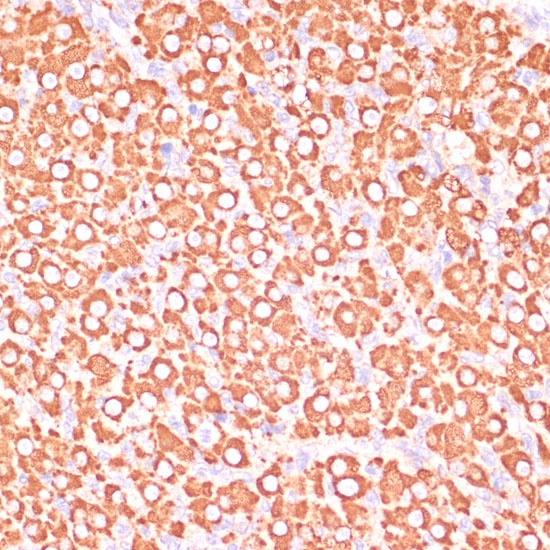
Immunohistochemistry analysis of paraffin-embedded Rat ovary using MEK3 Rabbit pAb (CAB7718) at dilution of 1:100 (40x lens). Microwave antigen retrieval performed with 0.01M PBS Buffer (pH 7.2) prior to IHC staining.
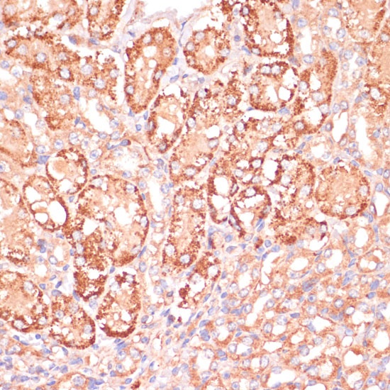
Immunohistochemistry analysis of paraffin-embedded Mouse kidney using MEK3 Rabbit pAb (CAB7718) at dilution of 1:100 (40x lens). Microwave antigen retrieval performed with 0.01M PBS Buffer (pH 7.2) prior to IHC staining.
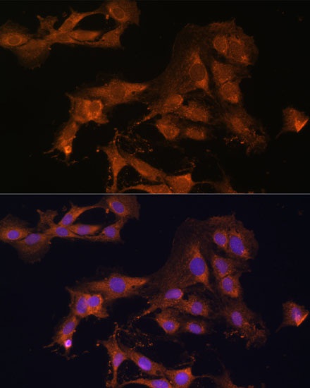
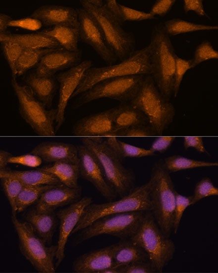
Immunofluorescence analysis of C6 cells using MEK3 Rabbit pAb (CAB7718) at dilution of 1:100. Secondary antibody: Cy3-conjugated Goat anti-Rabbit IgG (H+L) (CABS007) at 1:500 dilution. Blue: DAPI for nuclear staining.
Immunofluorescence analysis of U-2 OS cells using MEK3 Rabbit pAb (CAB7718) at dilution of 1:100. Secondary antibody: Cy3-conjugated Goat anti-Rabbit IgG (H+L) (CABS007) at 1:500 dilution. Blue: DAPI for nuclear staining.