The [KO Validated] MAP3K1 Antibody (CAB18041) is a high-quality antibody developed for reliable detection and analysis of target proteins. This antibody, generated in rabbits, has been validated for use in various experimental applications, particularly Western blot analysis. It specifically binds to the MAP3K1 protein, enabling precise detection and characterization in different cell types.MAP3K1 is a crucial component of signaling cascades involved in cell growth, differentiation, and survival. Dysregulation of MAP3K1 activity has been linked to various diseases, including cancer, making it a promising target for therapeutic intervention.
This antibody is validated for use in WB, IHC-P, ELISA applications and has demonstrated reactivity against Human, Mouse, Rat samples.
Product Name:
[KO Validated] MAP3K1 Antibody
SKU:
CAB18041
Size:
20μL, 100μL
Reactivity:
Human, Mouse, Rat
Conjugate:
Unconjugated
Immunogen:
Synthetic peptide. This information is considered to be commercially sensitive.
Recommended starting concentration is 1 μg/mL. Please optimize the concentration based on your specific assay requirements.
Synonyms:
MEKK, MEKK1, SRXY6, MEKK 1, MAPKKK1, K1
Positive Sample:
HeLa, Raji, A-431
Cellular Localization:
Cytoplasm, Cytosol.
Calculated MW:
164kDa
Observed MW:
164kDa
The protein encoded by this gene is a serine/threonine kinase and is part of some signal transduction cascades, including the ERK and JNK kinase pathways as well as the NF-kappa-B pathway. The encoded protein is activated by autophosphorylation and requires magnesium as a cofactor in phosphorylating other proteins. This protein has E3 ligase activity conferred by a plant homeodomain (PHD) in its N-terminus and phospho-kinase activity conferred by a kinase domain in its C-terminus.
Purification Method
Affinity purification
Gene ID
4214
RRID
AB_2861837
Buffer Information
Store at -20℃. Avoid freeze / thaw cycles. Buffer: PBS with 0.01% thimerosal,50% glycerol,pH7.3.
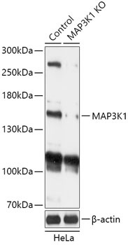
Western blot analysis of lysates from wild type (WT) and MAP3K1 knockout (KO) HeLa cells, using [KO Validated] MAP3K1 Rabbit pAb (CAB18041) at 1:1000 dilution. Secondary antibody: HRP-conjugated Goat anti-Rabbit IgG (H+L) (CABS014) at 1:10000 dilution. Lysates/proteins: 25μg per lane. Blocking buffer: 3% nonfat dry milk in TBST. Detection: ECL Basic Kit (AbGn00020). Exposure time: 1min.
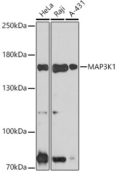
Western blot analysis of various lysates using MAP3K1 (CAB18041) at 1:1000 dilution. Secondary antibody: HRP-conjugated Goat anti-Rabbit IgG (H+L) (CABS014) at 1:10000 dilution. Lysates/proteins: 25μg per lane. Blocking buffer: 3% nonfat dry milk in TBST. Detection: ECL Basic Kit (AbGn00020). Exposure time: 30s.
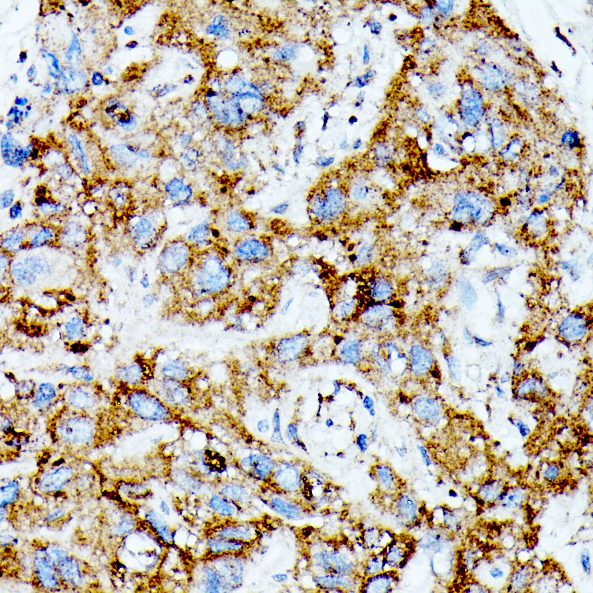
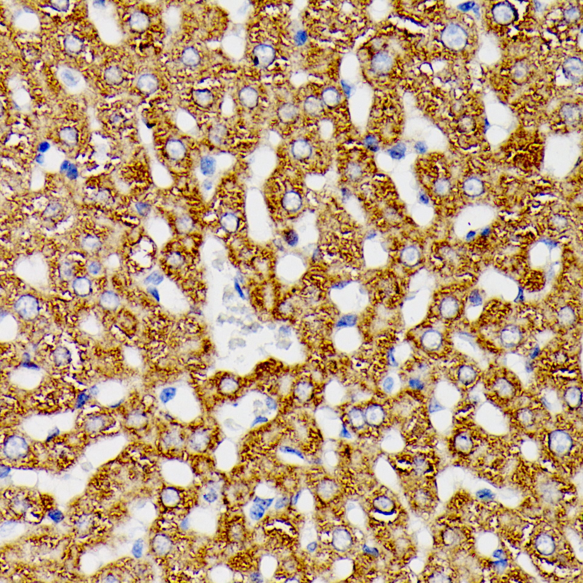
Immunohistochemistry analysis of paraffin-embedded Human liver cancer tissue using MAP3K1 Rabbit pAb (CAB18041) at a dilution of 1:100 (40x lens). Microwave antigen retrieval was performed with 0.01 M Tris-EDTA repair solution (pH 9.0) prior to IHC staining.
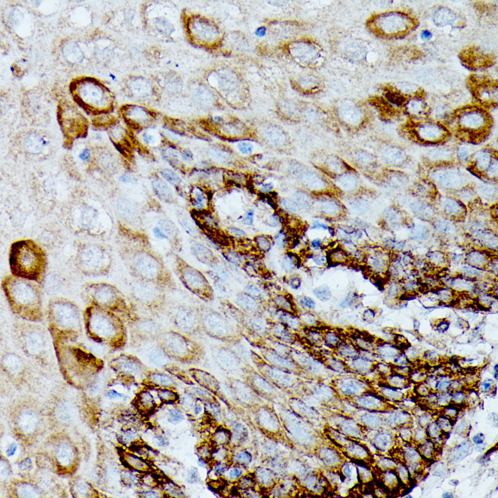
Immunohistochemistry analysis of paraffin-embedded Human esophagus tissue using MAP3K1 Rabbit pAb (CAB18041) at a dilution of 1:100 (40x lens). Microwave antigen retrieval was performed with 0.01 M Tris-EDTA repair solution (pH 9.0) prior to IHC staining.
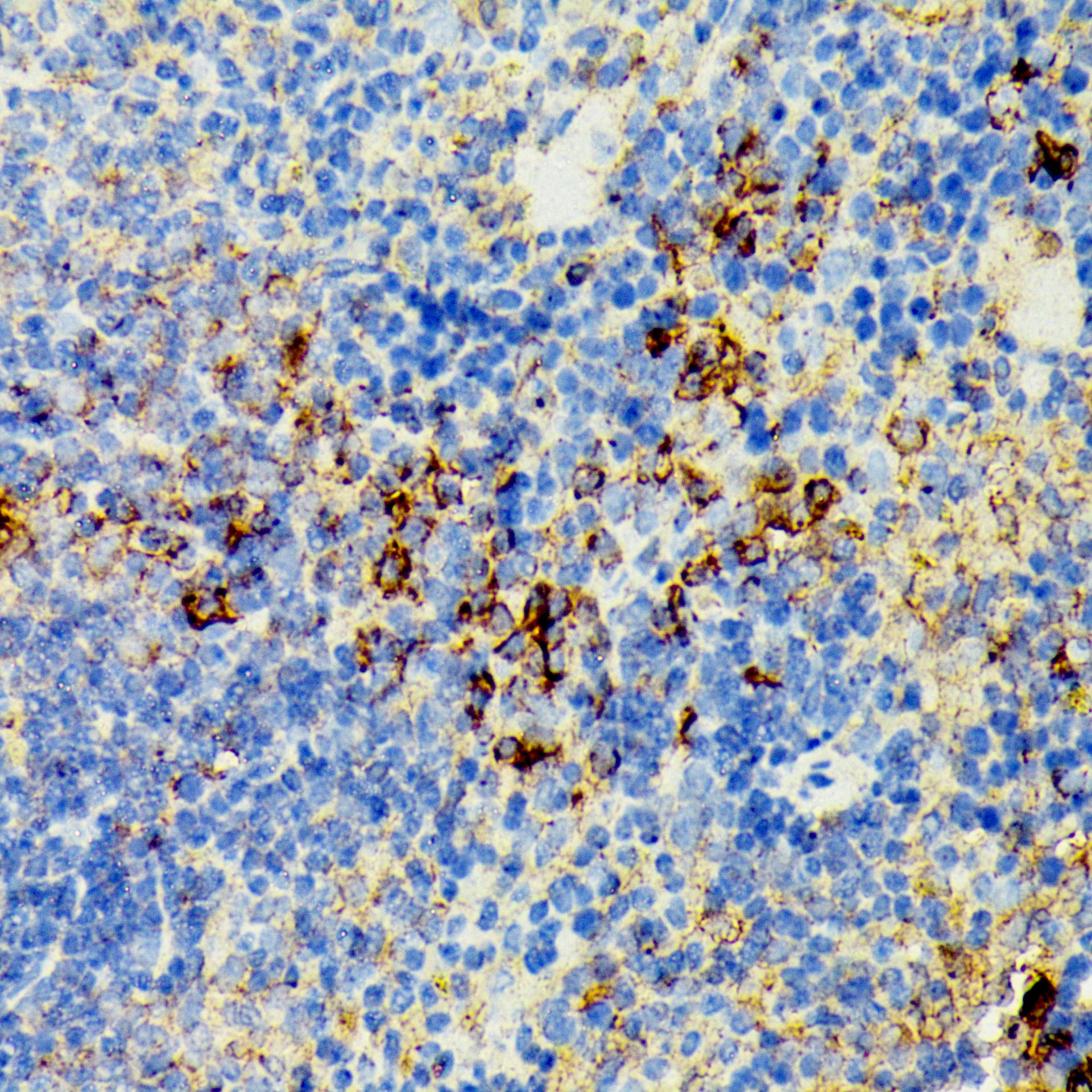
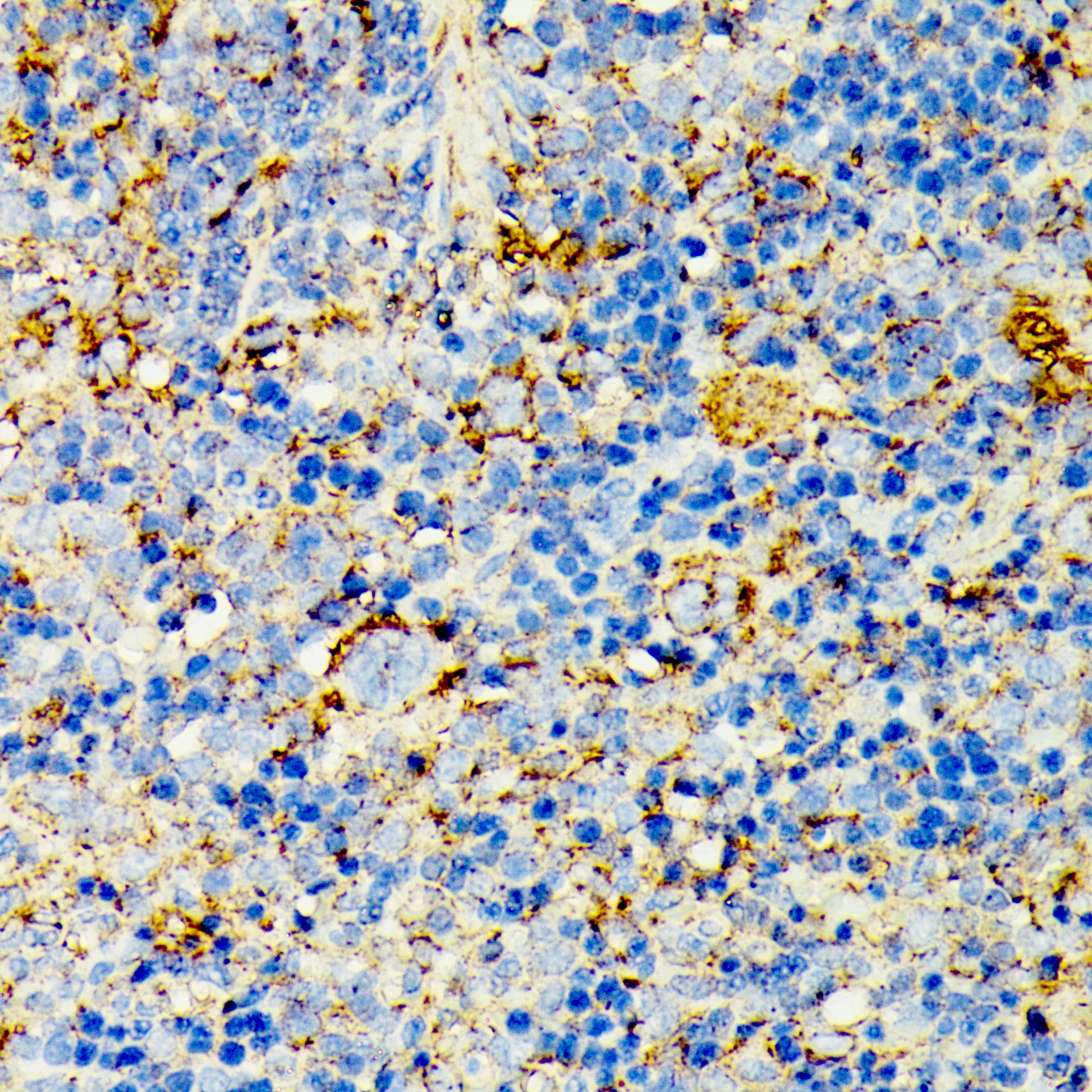
Immunohistochemistry analysis of paraffin-embedded Mouse spleen tissue using MAP3K1 Rabbit pAb (CAB18041) at a dilution of 1:100 (40x lens). Microwave antigen retrieval was performed with 0.01 M Tris-EDTA repair solution (pH 9.0) prior to IHC staining.
Immunohistochemistry analysis of paraffin-embedded Rat spleen tissue using MAP3K1 Rabbit pAb (CAB18041) at a dilution of 1:100 (40x lens). Microwave antigen retrieval was performed with 0.01 M Tris-EDTA repair solution (pH 9.0) prior to IHC staining.
Immunohistochemistry analysis of paraffin-embedded Rat liver tissue using MAP3K1 Rabbit pAb (CAB18041) at a dilution of 1:100 (40x lens). Microwave antigen retrieval was performed with 0.01 M Tris-EDTA repair solution (pH 9.0) prior to IHC staining.