The ERK5 Antibody (CAB2111) is a high-quality antibody developed for reliable detection and analysis of target proteins. This antibody, raised in rabbits, exhibits high reactivity with human samples and has been validated for use in Western blot applications.MAPK7, also known as ERK5, is a key signaling molecule involved in cell growth and survival pathways. Its dysregulation has been linked to various diseases, including cancer, cardiovascular disorders, and neurodegenerative diseases.
This antibody is validated for use in WB, IHC-P, IF/ICC, ELISA applications and has demonstrated reactivity against Human, Mouse, Rat samples.
Product Name:
ERK5 Antibody
SKU:
CAB2111
Size:
20μL, 100μL
Reactivity:
Human, Mouse, Rat
Conjugate:
Unconjugated
Immunogen:
Recombinant protein (or fragment).This information is considered to be commercially sensitive.
Recommended starting concentration is 1 μg/mL. Please optimize the concentration based on your specific assay requirements.
Synonyms:
BMK1, ERK4, ERK5, PRKM7
Positive Sample:
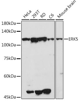
HeLa, 293T, RD, C6, Mouse brain
Cellular Localization:
Cytoplasm, Nucleus, Pml Body.
Calculated MW:
88kDa
Observed MW:
115kDa
The protein encoded by this gene is a member of the MAP kinase family. MAP kinases act as an integration point for multiple biochemical signals, and are involved in a wide variety of cellular processes such as proliferation, differentiation, transcription regulation and development. This kinase is specifically activated by mitogen-activated protein kinase kinase 5 (MAP2K5/MEK5). It is involved in the downstream signaling processes of various receptor molecules including receptor type kinases, and G protein-coupled receptors. In response to extracelluar signals, this kinase translocates to cell nucleus, where it regulates gene expression by phosphorylating, and activating different transcription factors. Four alternatively spliced transcript variants of this gene encoding two distinct isoforms have been reported.
Purification Method
Affinity purification
Gene ID
5598
RRID
AB_2764130
Buffer Information
Store at -20℃. Avoid freeze / thaw cycles. Buffer: PBS containing 50% glycerol, preserved with proclin300 or sodium azide, pH 7.3.
Western blot analysis of various lysates using ERK5 Rabbit pAb (CAB2111) at 1:500 dilution. Secondary antibody: HRP-conjugated Goat anti-Rabbit IgG (H+L) (CABS014) at 1:10000 dilution. Lysates/proteins: 25μg per lane. Blocking buffer: 3% nonfat dry milk in TBST. Detection: ECL Basic Kit (AbGn00020). Exposure time: 90s.
Immunohistochemistry analysis of paraffin-embedded Mouse lung using ERK5 Rabbit pAb (CAB2111) at dilution of 1:50 (40x lens). High pressure antigen retrieval performed with 0.01M Citrate buffer (pH 6.0) prior to IHC staining.
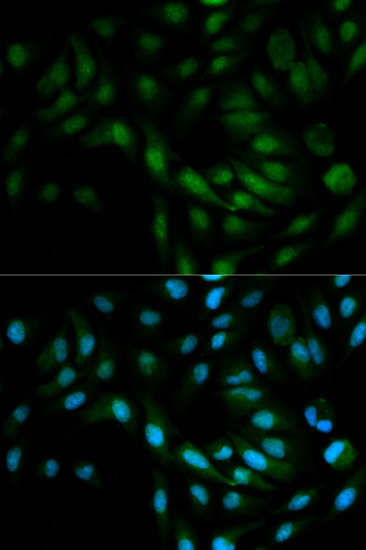
Immunofluorescence analysis of MCF-7 cells using ERK5 Rabbit pAb (CAB2111). Secondary antibody: Cy3-conjugated Goat anti-Rabbit IgG (H+L) (CABS007) at 1:500 dilution. Blue: DAPI for nuclear staining.