The MAPKAPK-2/MK2 Monoclonal Antibody (CAB22183) is a high-quality antibody developed for reliable detection and analysis of target proteins. This antibody, generated through monoclonal techniques, specifically targets the MK2 protein in human samples and is validated for use in a variety of applications, including Western blot and immunohistochemistry.MAPKAPK-2, also known as mitogen-activated protein kinase-activated protein kinase 2, plays a crucial role in regulating cell survival, proliferation, and differentiation in response to various stimuli. Its involvement in inflammatory processes and stress signaling makes it a promising target for studying diseases such as cancer, neurodegenerative disorders, and inflammatory conditions.
This antibody is validated for use in WB, ELISA applications and has demonstrated reactivity against Human, Mouse, Rat samples.
Product Name:
MAPKAPK-2/MK2 Monoclonal Antibody
SKU:
CAB22183
Size:
20μL, 100μL
Reactivity:
Human, Mouse, Rat
Clone Number:
ARC55427
Conjugate:
Unconjugated
Immunogen:
Synthetic peptide. This information is considered to be commercially sensitive.
Recommended starting concentration is 1 μg/mL. Please optimize the concentration based on your specific assay requirements.
Synonyms:
MK2, MK-2, MAPKAP-K2, MAPKAPK-2/MK2
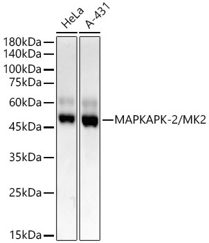
Positive Sample:
HeLa, A-431
Cellular Localization:
Cytoplasm, Nucleus.
Calculated MW:
46kDa
Observed MW:
47kDa
This gene encodes a member of the Ser/Thr protein kinase family. This kinase is regulated through direct phosphorylation by p38 MAP kinase. In conjunction with p38 MAP kinase, this kinase is known to be involved in many cellular processes including stress and inflammatory responses, nuclear export, gene expression regulation and cell proliferation. Heat shock protein HSP27 was shown to be one of the substrates of this kinase in vivo. Two transcript variants encoding two different isoforms have been found for this gene.
Purification Method
Affinity purification
Gene ID
9261
Buffer Information
Store at -20℃. Avoid freeze / thaw cycles. Buffer: PBS with 0.09% Sodium azide,0.05% BSA,50% glycerol,pH7.3.
Western blot analysis of various lysates, using MAPKAPK-2/MK2 Rabbit mAb (CAB22183) at1:2000 dilution. Secondary antibody: HRP-conjugated Goat anti-Rabbit IgG (H+L) (CABS014) at 1:10000 dilution. Lysates/proteins: 25μg per lane. Blocking buffer: 3% nonfat dry milk in TBST. Detection: ECL Basic Kit (AbGn00020). Exposure time: 30s.
 at1:2000 dilution. Secondary antibody: HRP Goat Anti-Rabbit IgG (H+L) at 1:10000 dilution. Lysates/proteins: 25μg per lane. Blocking buffer: 3% nonfat dry milk in TBST.")
 at1:2000 dilution. Secondary antibody: HRP Goat Anti-Rabbit IgG (H+L) at 1:10000 dilution. Lysates/proteins: 25μg per lane. Blocking buffer: 3% nonfat dry milk in TBST.")

 at 1:10000 dilution. Lysates/proteins: 25ug per lane. Blocking buffer: 3% nonfat dry milk in TBST. Detection: ECL Basic Kit. Exposure time: 10s.")
