The MARCKS Antibody (CAB0936) is a high-quality antibody developed for reliable detection and analysis of target proteins. The protein encoded by this gene is a substrate for protein kinase C. It is localized to the plasma membrane and is an actin filament crosslinking protein. Phosphorylation by protein kinase C or binding to calcium-calmodulin inhibits its association with actin and with the plasma membrane, leading to its presence in the cytoplasm. The protein is thought to be involved in cell motility, phagocytosis, membrane trafficking and mitogenesis. RRID AB_2757467 Gene ID 4082 Swiss Prot Synonym MACS; 80K-L; PKCSL; PRKCSL; MARCKS
This antibody is validated for use in WB, IF/ICC, ELISA applications and has demonstrated reactivity against Human, Mouse, Rat samples.
Product Name:
MARCKS Antibody
SKU:
CAB0936
Size:
100μL, 20μL
Reactivity:
Human, Mouse, Rat
Clone Number:
-
Conjugate:
Unconjugated
Immunogen:
Synthetic peptide. This information is considered to be commercially sensitive.
Tested Applications:
WBIF/ICCELISA
Recommended Dilution:
WB
1:500 - 1:2000
IF
/
ICC
1:50 - 1:200
ELISA
Recommended starting concentration is 1 μg/mL. Please optimize the concentration based on your specific assay requirements.
Synonyms:
MACS, 80K-L, PKCSL, PRKCSL, MARCKS
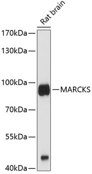
Positive Sample:
Rat brain
Cellular Localization:
Cytoplasm, Lipid-Anchor, Membrane, Cytoskeleton.
Calculated MW:
32kDa
Observed MW:
80kDa
The protein encoded by this gene is a substrate for protein kinase C. It is localized to the plasma membrane and is an actin filament crosslinking protein. Phosphorylation by protein kinase C or binding to calcium-calmodulin inhibits its association with actin and with the plasma membrane, leading to its presence in the cytoplasm. The protein is thought to be involved in cell motility, phagocytosis, membrane trafficking and mitogenesis. RRID AB_2757467 Gene ID 4082 Swiss Prot Synonym MACS; 80K-L; PKCSL; PRKCSL; MARCKS
Purification Method:
Affinity purification
Gene ID:
4082
RRID:
AB_2757467
Buffer Information:
Store at -20℃. Avoid freeze / thaw cycles. Buffer: PBS containing 50% glycerol, preserved with proclin300 or sodium azide, pH 7.3.
Western blot analysis of lysates from rat brain, using MARCKS Rabbit pAb (CAB0936) at 1:1000 dilution. Secondary antibody: HRP-conjugated Goat anti-Rabbit IgG (H+L) (AS014) at 1:10000 dilution. Lysates/proteins: 25μg per lane. Blocking buffer: 3% nonfat dry milk in TBST. Detection: ECL Basic Kit (AbGn00020). Exposure time: 30s.
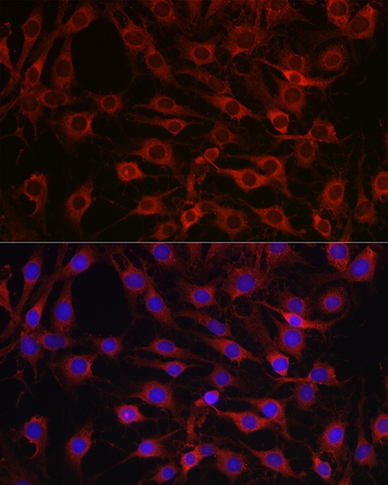
Immunofluorescence analysis of C6 cells using MARCKS Rabbit pAb (CAB0936) at dilution of 1:100 (40x lens). Secondary antibody: Cy3-conjugated Goat anti-Rabbit IgG (H+L) (AS007) at 1:500 dilution. Blue: DAPI for nuclear staining.
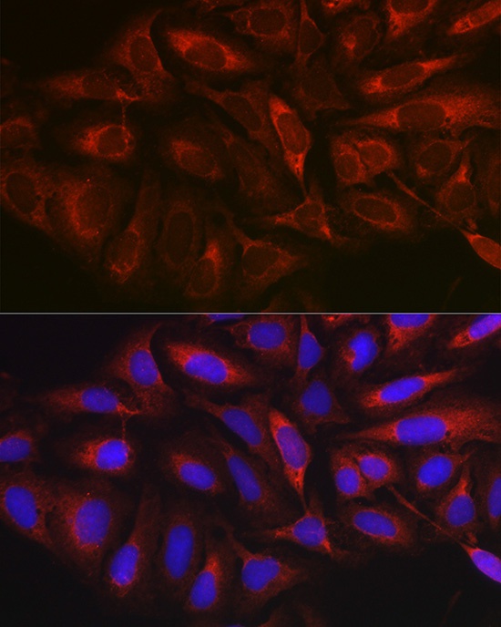
Immunofluorescence analysis of U-2 OS cells using MARCKS Rabbit pAb (CAB0936) at dilution of 1:100 (40x lens). Secondary antibody: Cy3-conjugated Goat anti-Rabbit IgG (H+L) (AS007) at 1:500 dilution. Blue: DAPI for nuclear staining.