The MBNL1 Monoclonal Antibody (CAB5149) is a high-quality antibody developed for reliable detection and analysis of target proteins. MBNL1 is known for its role in regulating alternative splicing of pre-mRNA molecules, particularly in the context of myotonic dystrophy, a neuromuscular disorder. This antibody, produced in rabbits, has been validated for use in Western blot and immunofluorescence applications, enabling the detection and visualization of MBNL1 in different cell types and tissues.MBNL1's involvement in RNA processing and splicing makes it a crucial player in the development and progression of diseases like myotonic dystrophy and certain types of cancer.
This antibody is validated for use in WB, IF/ICC, ELISA applications and has demonstrated reactivity against Human samples.
Product Name:
MBNL1 Monoclonal Antibody
SKU:
CAB5149
Size:
20μL, 100μL
Reactivity:
Human
Clone Number:
ARC1199
Conjugate:
Unconjugated
Immunogen:
Synthetic peptide. This information is considered to be commercially sensitive.
Recommended starting concentration is 1 μg/mL. Please optimize the concentration based on your specific assay requirements.
Synonyms:
EXP, MBNL, MBNL1
Positive Sample:
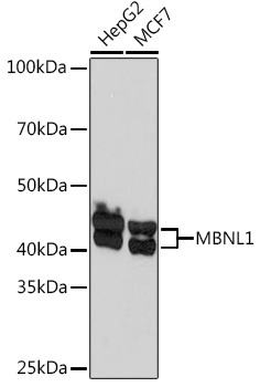
HepG2, MCF7
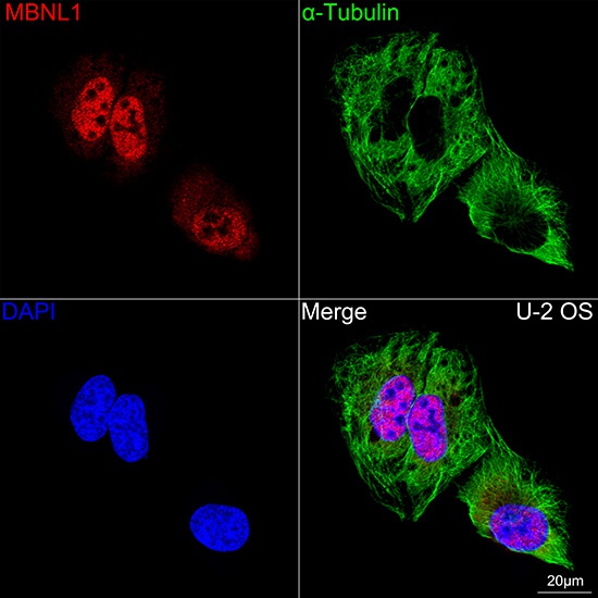
Cellular Localization:
Cytoplasm, Cytoplasmic Granule, Nucleus.
Calculated MW:
42kDa
Observed MW:
40kDa/42kDa
This gene encodes a member of the muscleblind protein family which was initially described in Drosophila melanogaster. The encoded protein is a C3H-type zinc finger protein that modulates alternative splicing of pre-mRNAs. Muscleblind proteins bind specifically to expanded dsCUG RNA but not to normal size CUG repeats and may thereby play a role in the pathophysiology of myotonic dystrophy. Mice lacking this gene exhibited muscle abnormalities and cataracts. Several alternatively spliced transcript variants have been described but the full-length natures of only some have been determined. The different isoforms are thought to have different binding specificities and/or splicing activities.
Purification Method
Affinity purification
Gene ID
4154
RRID
AB_2863467
Buffer Information
Store at -20℃. Avoid freeze / thaw cycles. Buffer: PBS containing 50% glycerol and 0.05% BSA, preserved with proclin300 or sodium azide, pH 7.3.
Western blot analysis of various lysates using MBNL1 Rabbit mAb (CAB5149) at 1:1000 dilution. Secondary antibody: HRP-conjugated Goat anti-Rabbit IgG (H+L) (CABS014) at 1:10000 dilution. Lysates/proteins: 25μg per lane. Blocking buffer: 3% nonfat dry milk in TBST. Detection: ECL Basic Kit (AbGn00020). Exposure time: 60s.
Confocal imaging of U-2 OS cells using MBNL1 Rabbit mAb (CAB5149, dilution 1:100) followed by a further incubation with Cy3 Goat Anti-Rabbit IgG (H+L) (CABS007, dilution 1:500) (Red). The cells were counterstained with α-Tubulin Mouse mAb (AC012, dilution 1:400) followed by incubation with ABflo® 488-conjugated Goat Anti-Mouse IgG (H+L) Ab (CABS076, dilution 1:500) (Green). DAPI was used for nuclear staining (Blue). Objective: 100x.