The MCM5 Antibody (CAB13514) is a high-quality antibody developed for reliable detection and analysis of target proteins. This antibody, generated in rabbits, exhibits high reactivity with human samples and has been validated for use in Western blot applications. By specifically binding to the MCM5 protein, this antibody enables precise detection and analysis in a variety of cell types, making it ideal for investigations in cell biology and cancer research.
This antibody is validated for use in WB, IHC-P, IF/ICC, IP, ELISA applications and has demonstrated reactivity against Human, Mouse, Rat samples.
Product Name:
MCM5 Antibody
SKU:
CAB13514
Size:
20μL, 100μL
Reactivity:
Human, Mouse, Rat
Conjugate:
Unconjugated
Immunogen:
Recombinant protein (or fragment).This information is considered to be commercially sensitive.
0.5μg-4μg antibody for 200μg-400μg extracts of whole cells
ELISA
Recommended starting concentration is 1 μg/mL. Please optimize the concentration based on your specific assay requirements.
Synonyms:
CDC46, MGORS8, P1-CDC46, MCM5
Positive Sample:
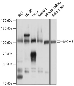
Raji, HL-60, HeLa, SW620, Mouse kidney, Rat kidney
Cellular Localization:
Nucleus.
Calculated MW:
82kDa
Observed MW:
100kDa
The protein encoded by this gene is structurally very similar to the CDC46 protein from S. cerevisiae, a protein involved in the initiation of DNA replication. The encoded protein is a member of the MCM family of chromatin-binding proteins and can interact with at least two other members of this family. The encoded protein is upregulated in the transition from the G0 to G1/S phase of the cell cycle and may actively participate in cell cycle regulation.
Purification Method
Affinity purification
Gene ID
4174
RRID
AB_2760376
Buffer Information
Store at -20℃. Avoid freeze / thaw cycles. Buffer: PBS containing 50% glycerol, preserved with proclin300 or sodium azide, pH 7.3.
Western blot analysis of various lysates using MCM5 Rabbit pAb (CAB13514) at 1:1000 dilution. Secondary antibody: HRP-conjugated Goat anti-Rabbit IgG (H+L) (CABS014) at 1:10000 dilution. Lysates/proteins: 25μg per lane. Blocking buffer: 3% nonfat dry milk in TBST. Detection: ECL Basic Kit (AbGn00020). Exposure time: 1s.
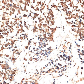
Immunohistochemistry analysis of paraffin-embedded Human lung cancer using MCM5 Rabbit pAb (CAB13514) at dilution of 1:100 (40x lens). Microwave antigen retrieval performed with 0.01M PBS Buffer (pH 7.2) prior to IHC staining.
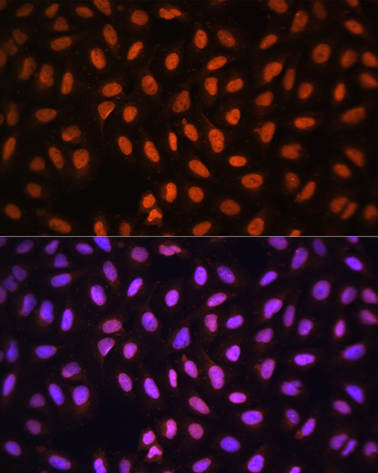
Immunofluorescence analysis of U-2 OS cells using MCM5 Rabbit pAb (CAB13514) at dilution of 1:100 (40x lens). Secondary antibody: Cy3-conjugated Goat anti-Rabbit IgG (H+L) (CABS007) at 1:500 dilution. Blue: DAPI for nuclear staining.
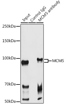
Immunoprecipitation analysis of 300 μg extracts of Raji cells using 3 μg MCM5 antibody (CAB13514). Western blot was performed from the immunoprecipitate using MCM5 antibody (CAB13514) at a dilution of 1:1000.