The MCU Antibody (CAB16281) is a high-quality antibody developed for reliable detection and analysis of target proteins. Enables calcium channel activity; identical protein binding activity; and uniporter activity. Involved in several processes, including positive regulation of mitochondrial calcium ion concentration; positive regulation of mitochondrial fission; and positive regulation of neutrophil chemotaxis. Acts upstream of or within calcium import into the mitochondrion. Located in mitochondrial inner membrane. Is integral component of mitochondrial inner membrane. Part of uniplex complex.
This antibody is validated for use in WB, IF/ICC, ELISA applications and has demonstrated reactivity against Human, Mouse, Rat samples.
Product Name:
MCU Antibody
SKU:
CAB16281
Size:
100μL, 20μL
Reactivity:
Human, Mouse, Rat
Conjugate:
Unconjugated
Immunogen:
Recombinant protein (or fragment).This information is considered to be commercially sensitive.
Tested Applications:
WBIF/ICCELISA
Recommended Dilution:
WB
1:1000 - 1:5000
IF/ICC
1:50 - 1:200
ELISA
Recommended starting concentration is 1 μg/mL. Please optimize the concentration based on your specific assay requirements.
Synonyms:
HsMCU, C10orf42, CCDC109A, MCU
Positive Sample:
HeLa
Cellular Localization:
Mitochondrial Inner Membrane, Mitochondrion.
Calculated MW:
40kDa
Observed MW:
35kDa
Enables calcium channel activity; identical protein binding activity; and uniporter activity. Involved in several processes, including positive regulation of mitochondrial calcium ion concentration; positive regulation of mitochondrial fission; and positive regulation of neutrophil chemotaxis. Acts upstream of or within calcium import into the mitochondrion. Located in mitochondrial inner membrane. Is integral component of mitochondrial inner membrane. Part of uniplex complex.
Purification Method
Affinity purification
Gene ID
90550
RRID
AB_2770322
Buffer Information
Store at -20℃. Avoid freeze / thaw cycles. Buffer: PBS containing 50% glycerol, preserved with proclin300 or sodium azide, pH 7.3.
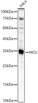
Western blot analysis of lysates from HeLa cells using MCU Rabbit pAb (CAB16281) at 1:2000 dilution. Secondary antibody: HRP-conjugated Goat anti-Rabbit IgG (H+L) (AS014) at 1:10000 dilution. Lysates/proteins: 25 μg per lane. Blocking buffer: 3% nonfat dry milk in TBST. Detection: ECL Basic Kit (AbGn00020). Exposure time: 30s.
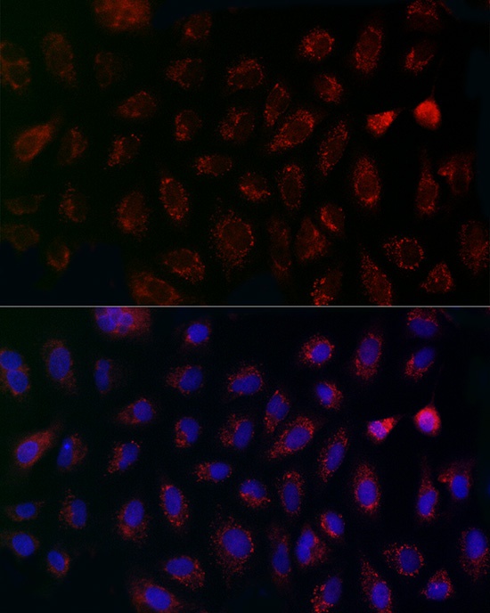
Immunofluorescence analysis of A-549 cells using MCU Rabbit pAb (CAB16281) at dilution of 1:200 (40x lens). Secondary antibody: Cy3-conjugated Goat anti-Rabbit IgG (H+L) (AS007) at 1:500 dilution. Blue: DAPI for nuclear staining.
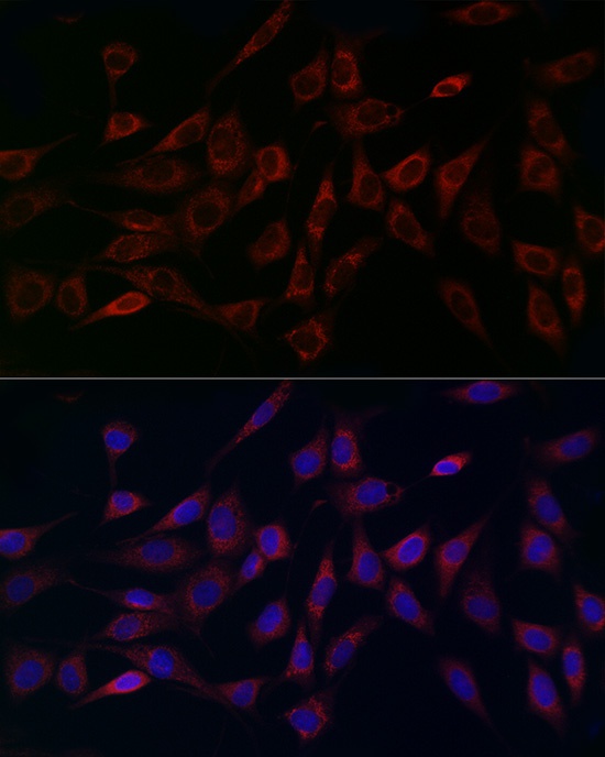
Immunofluorescence analysis of NIH/3T3 cells using MCU Rabbit pAb (CAB16281) at dilution of 1:200 (40x lens). Secondary antibody: Cy3-conjugated Goat anti-Rabbit IgG (H+L) (AS007) at 1:500 dilution. Blue: DAPI for nuclear staining.
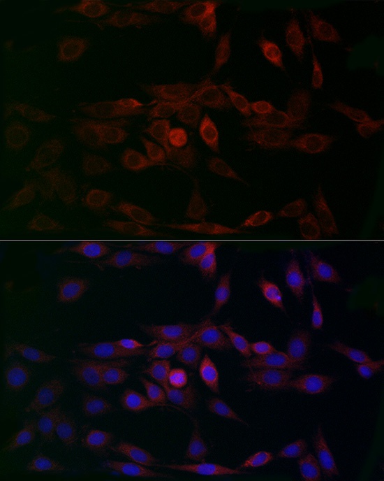
Immunofluorescence analysis of PC-12 cells using MCU Rabbit pAb (CAB16281) at dilution of 1:200 (40x lens). Secondary antibody: Cy3-conjugated Goat anti-Rabbit IgG (H+L) (AS007) at 1:500 dilution. Blue: DAPI for nuclear staining.