The MDH1 Antibody (CAB7563) is a high-quality antibody developed for reliable detection and analysis of target proteins. This antibody, produced in rabbits, exhibits high reactivity with human samples and has been validated for use in Western blot applications.MDH1, also known as malate dehydrogenase 1, plays a crucial role in converting malate to oxaloacetate, a key step in the citric acid cycle that generates energy for cellular processes. Dysregulation of MDH1 has been implicated in various diseases, including cancer and metabolic disorders, making it an important target for research.
This antibody is validated for use in WB, ELISA applications and has demonstrated reactivity against Human, Mouse, Rat samples.
Product Name:
MDH1 Antibody
SKU:
CAB7563
Size:
20μL, 100μL
Reactivity:
Human, Mouse, Rat
Conjugate:
Unconjugated
Immunogen:
Recombinant protein (or fragment).This information is considered to be commercially sensitive.
This gene encodes an enzyme that catalyzes the NAD/NADH-dependent, reversible oxidation of malate to oxaloacetate in many metabolic pathways, including the citric acid cycle. Two main isozymes are known to exist in eukaryotic cells: one is found in the mitochondrial matrix and the other in the cytoplasm. This gene encodes the cytosolic isozyme, which plays a key role in the malate-aspartate shuttle that allows malate to pass through the mitochondrial membrane to be transformed into oxaloacetate for further cellular processes. Alternatively spliced transcript variants have been found for this gene. A recent study showed that a C-terminally extended isoform is produced by use of an alternative in-frame translation termination codon via a stop codon readthrough mechanism, and that this isoform is localized in the peroxisomes. Pseudogenes have been identified on chromosomes X and 6.
Purification Method
Affinity purification
Gene ID
4190
RRID
AB_2768088
Buffer Information
Store at -20℃. Avoid freeze / thaw cycles. Buffer: PBS containing 50% glycerol, preserved with proclin300 or sodium azide, pH 7.3.
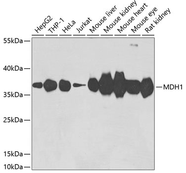
Western blot analysis of various lysates using MDH1 Rabbit pAb (CAB7563) at 1:3000 dilution. Secondary antibody: HRP-conjugated Goat anti-Rabbit IgG (H+L) (CABS014) at 1:10000 dilution. Lysates/proteins: 25μg per lane. Blocking buffer: 3% nonfat dry milk in TBST. Detection: ECL Basic Kit (AbGn00020). Exposure time: 90s.