The MDM2 Antibody (CAB0345) is a high-quality antibody developed for reliable detection and analysis of target proteins. This antibody, generated in rabbits, shows high reactivity with human samples and is specifically validated for use in Western blotting applications. By targeting the MDM2 protein, this antibody enables precise detection and analysis in a variety of cell types, making it an ideal choice for studies in oncology and molecular biology.MDM2, also known as mouse double minute 2 homolog, is a critical player in controlling the activity of the tumor suppressor protein p53. Dysregulation of the MDM2-p53 pathway is implicated in numerous cancers, making MDM2 an attractive target for cancer therapy research.
This antibody is validated for use in WB, IHC-P, ELISA applications and has demonstrated reactivity against Human, Mouse samples.
Product Name:
MDM2 Antibody
SKU:
CAB0345
Size:
20μL, 100μL
Reactivity:
Human, Mouse
Conjugate:
Unconjugated
Immunogen:
Synthetic peptide. This information is considered to be commercially sensitive.
Recommended starting concentration is 1 μg/mL. Please optimize the concentration based on your specific assay requirements.
Synonyms:
HDMX, LSKB, hdm2, ACTFS, MDM2
Positive Sample:
NIH/3T3 treated with MG132
Cellular Localization:
Cytoplasm, Nucleus, Nucleolus, Nucleoplasm.
Calculated MW:
55kDa
Observed MW:
90kDa
This gene encodes a nuclear-localized E3 ubiquitin ligase. The encoded protein can promote tumor formation by targeting tumor suppressor proteins, such as p53, for proteasomal degradation. This gene is itself transcriptionally-regulated by p53. Overexpression or amplification of this locus is detected in a variety of different cancers. There is a pseudogene for this gene on chromosome 2. Alternative splicing results in a multitude of transcript variants, many of which may be expressed only in tumor cells.
Purification Method
Affinity purification
Gene ID
4193
RRID
AB_2757138
Buffer Information
Store at -20℃. Avoid freeze / thaw cycles. Buffer: PBS containing 50% glycerol, preserved with proclin300 or sodium azide, pH 7.3.
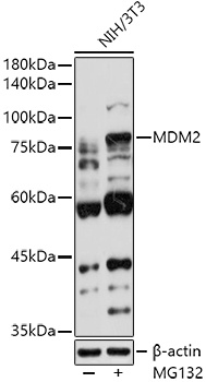
Western blot analysis of lysates from NIH/3T3 cells, using MDM2 Rabbit pAb (CAB0345) at 1:1000 dilution. NIH/3T3 cells were treated with MG132(50 μM) at 37℃ for 90 minutes. Secondary antibody: HRP-conjugated Goat anti-Rabbit IgG (H+L) (CABS014) at 1:10000 dilution. Lysates/proteins: 25μg per lane. Blocking buffer: 3% nonfat dry milk in TBST. Detection: ECL Basic Kit (AbGn00020). Exposure time: 90s.
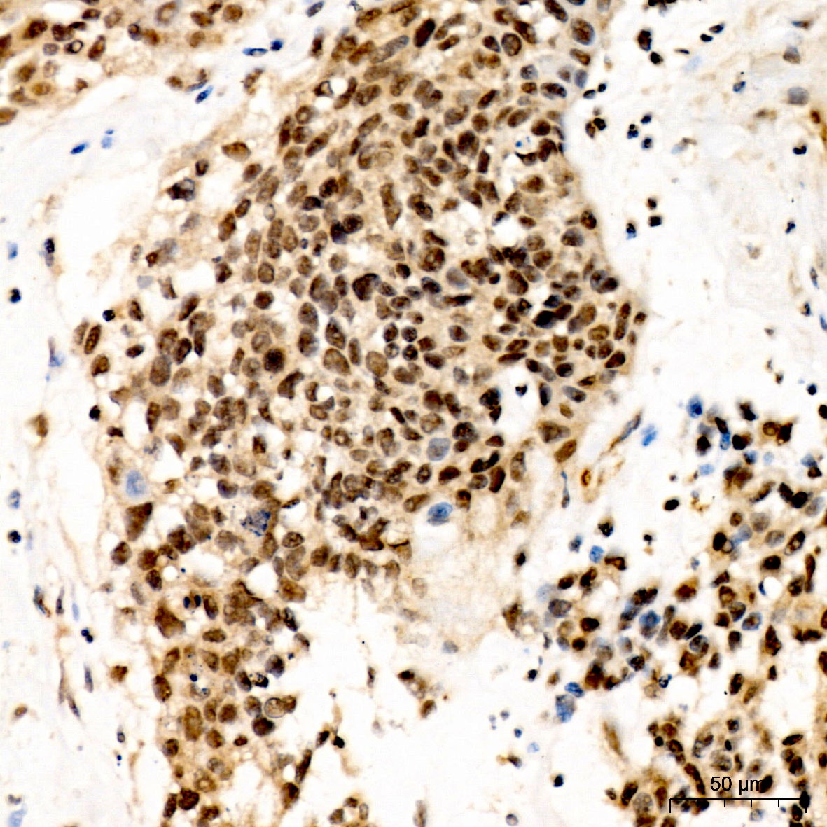
Immunohistochemistry analysis of paraffin-embedded Human urothelial carcinoma using MDM2 Rabbit pAb (CAB0345) at dilution of 1:50 (40x lens). High pressure antigen retrieval performed with 0.01M Citrate buffer (pH 6.0) prior to IHC staining.