The ME3 Antibody (CAB15787) is a high-quality antibody developed for reliable detection and analysis of target proteins. Malic enzyme catalyzes the oxidative decarboxylation of malate to pyruvate using either NAD+ or NADP+ as a cofactor. Mammalian tissues contain 3 distinct isoforms of malic enzyme: a cytosolic NADP(+)-dependent isoform, a mitochondrial NADP(+)-dependent isoform, and a mitochondrial NAD(+)-dependent isoform. This gene encodes a mitochondrial NADP(+)-dependent isoform. Multiple alternatively spliced transcript variants have been found for this gene, but the biological validity of some variants has not been determined.
This antibody is validated for use in WB, IHC-P, IF/ICC, ELISA applications and has demonstrated reactivity against Human, Mouse, Rat samples.
Product Name:
ME3 Antibody
SKU:
CAB15787
Size:
100μL, 20μL
Reactivity:
Human, Mouse, Rat
Conjugate:
Unconjugated
Immunogen:
Recombinant protein (or fragment).This information is considered to be commercially sensitive.
Tested Applications:
WBIHC-PIF/ICCELISA
Recommended Dilution:
WB
1:500 - 1:2000
IHC-P
1:50 - 1:200
IF/ICC
1:50 - 1:200
ELISA
Recommended starting concentration is 1 μg/mL. Please optimize the concentration based on your specific assay requirements.
Synonyms:
NADP-ME, ME3
Positive Sample:
HT-29, 293T, Mouse brain, Mouse heart, Mouse kidney, Rat brain, Rat heart, Rat kidney
Cellular Localization:
Mitochondrion Matrix.
Calculated MW:
67kDa
Observed MW:
78kDa
Malic enzyme catalyzes the oxidative decarboxylation of malate to pyruvate using either NAD+ or NADP+ as a cofactor. Mammalian tissues contain 3 distinct isoforms of malic enzyme: a cytosolic NADP(+)-dependent isoform, a mitochondrial NADP(+)-dependent isoform, and a mitochondrial NAD(+)-dependent isoform. This gene encodes a mitochondrial NADP(+)-dependent isoform. Multiple alternatively spliced transcript variants have been found for this gene, but the biological validity of some variants has not been determined.
Purification Method
Affinity purification
Gene ID
10873
RRID
AB_2763207
Buffer Information
Store at -20℃. Avoid freeze / thaw cycles. Buffer: PBS with 0.01% thimerosal,50% glycerol,pH7.3.
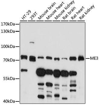
Western blot analysis of various lysates using ME3 Rabbit pAb (CAB15787) at 1:1000 dilution. Secondary antibody: HRP-conjugated Goat anti-Rabbit IgG (H+L) (AS014) at 1:10000 dilution. Lysates/proteins: 25μg per lane. Blocking buffer: 3% nonfat dry milk in TBST. Detection: ECL Basic Kit (AbGn00020). Exposure time: 3min.
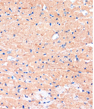
Immunohistochemistry analysis of paraffin-embedded Mouse heart using ME3 Rabbit pAb (CAB15787) at dilution of 1:100 (40x lens). Microwave antigen retrieval performed with 0.01M PBS Buffer (pH 7.2) prior to IHC staining.
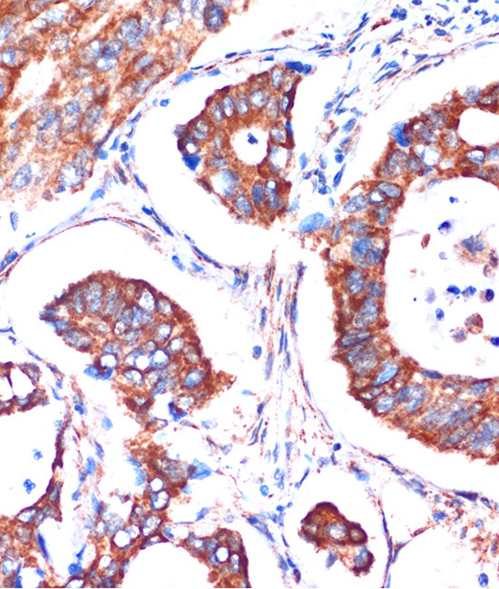
Immunohistochemistry analysis of paraffin-embedded Human colon carcinoma using ME3 Rabbit pAb (CAB15787) at dilution of 1:100 (40x lens). Microwave antigen retrieval performed with 0.01M PBS Buffer (pH 7.2) prior to IHC staining.
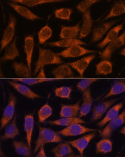
Immunofluorescence analysis of L929 cells using ME3 Rabbit pAb (CAB15787) at dilution of 1:100. Secondary antibody: Cy3-conjugated Goat anti-Rabbit IgG (H+L) (AS007) at 1:500 dilution. Blue: DAPI for nuclear staining.