The MKL1 Antibody (CAB8504) is a high-quality antibody developed for reliable detection and analysis of target proteins. This antibody is produced in rabbits and specifically recognizes MKL1 in human samples, making it an excellent choice for Western blot applications. By targeting the MKL1 protein, researchers can accurately detect and analyze its expression levels in various cell types, providing valuable insights into its role in cellular function and gene regulation.MKL1, also known as MRTF-A, is a key player in cellular processes such as migration, adhesion, and differentiation.
This antibody is validated for use in WB, IHC-P, IF/ICC, ELISA applications and has demonstrated reactivity against Human, Mouse, Rat samples.
Product Name:
MKL1 Antibody
SKU:
CAB8504
Size:
20μL, 100μL
Reactivity:
Human, Mouse, Rat
Conjugate:
Unconjugated
Immunogen:
Recombinant protein (or fragment).This information is considered to be commercially sensitive.
Recommended starting concentration is 1 μg/mL. Please optimize the concentration based on your specific assay requirements.
Synonyms:
MAL, MKL, BSAC, MKL1, MRTF-A
Positive Sample:
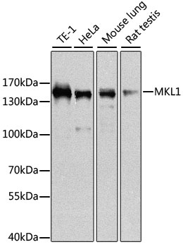
TE-1, HeLa, Mouse lung, Rat testis
Cellular Localization:
Cytoplasm, Nucleus.
Calculated MW:
99kDa
Observed MW:
145kDa
The protein encoded by this gene interacts with the transcription factor myocardin, a key regulator of smooth muscle cell differentiation. The encoded protein is predominantly nuclear and may help transduce signals from the cytoskeleton to the nucleus. This gene is involved in a specific translocation event that creates a fusion of this gene and the RNA-binding motif protein-15 gene. This translocation has been associated with acute megakaryocytic leukemia. Alternative splicing results in multiple transcript variants.
Purification Method
Affinity purification
Gene ID
57591
RRID
AB_2770370
Buffer Information
Store at -20℃. Avoid freeze / thaw cycles. Buffer: PBS containing 50% glycerol, preserved with proclin300 or sodium azide, pH 7.3.
Western blot analysis of various lysates using MKL1 Rabbit pAb (CAB8504) at 1:1000 dilution. Secondary antibody: HRP-conjugated Goat anti-Rabbit IgG (H+L) (CABS014) at 1:10000 dilution. Lysates/proteins: 25μg per lane. Blocking buffer: 3% nonfat dry milk in TBST. Detection: ECL Basic Kit (AbGn00020). Exposure time: 10s.
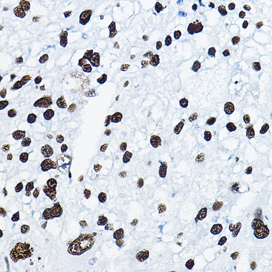
Immunohistochemistry analysis of paraffin-embedded Human liver cancer using MKL1 Rabbit pAb (CAB8504) at dilution of 1:100 (40x lens). Microwave antigen retrieval performed with 0.01M PBS Buffer (pH 7.2) prior to IHC staining.
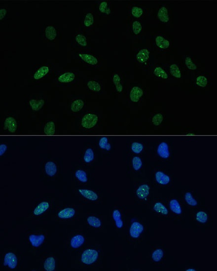
Immunofluorescence analysis of U-2 OS cells using MKL1 Rabbit pAb (CAB8504) at dilution of 1:100 (40x lens). Secondary antibody: Cy3-conjugated Goat anti-Rabbit IgG (H+L) (CABS007) at 1:500 dilution. Blue: DAPI for nuclear staining.