The MMP14/MT1-MMP Antibody (CAB2549) is a high-quality antibody developed for reliable detection and analysis of target proteins. Raised in rabbits, this antibody is highly specific and reactive with human samples, making it ideal for use in Western blot applications. By binding to the MMP14 protein, this antibody enables researchers to detect and analyze MMP14 expression in various cell types, aiding studies in cancer progression, metastasis, and tissue repair mechanisms.MMP14, also known as membrane-type matrix metalloproteinase 1 (MT1-MMP), plays a key role in regulating cell invasion, migration, and angiogenesis.
This antibody is validated for use in WB, IHC-P, IF/ICC, ELISA applications and has demonstrated reactivity against Human, Mouse, Rat samples.
Product Name:
MMP14/MT1-MMP Antibody
SKU:
CAB2549
Size:
20μL, 100μL
Reactivity:
Human, Mouse, Rat
Conjugate:
Unconjugated
Immunogen:
Synthetic peptide. This information is considered to be commercially sensitive.
Cytoplasm, Melanosome, Membrane, Single-Pass Type I Membrane Protein.
Calculated MW:
66kDa
Observed MW:
60kDa
Proteins of the matrix metalloproteinase (MMP) family are involved in the breakdown of extracellular matrix in normal physiological processes, such as embryonic development, reproduction, and tissue remodeling, as well as in disease processes, such as arthritis and metastasis. Most MMP's are secreted as inactive proproteins which are activated when cleaved by extracellular proteinases. However, the protein encoded by this gene is a member of the membrane-type MMP (MT-MMP) subfamily; each member of this subfamily contains a potential transmembrane domain suggesting that these proteins are expressed at the cell surface rather than secreted. This protein activates MMP2 protein, and this activity may be involved in tumor invasion.
Purification Method
Affinity purification
Gene ID
4323
RRID
AB_2764438
Buffer Information
Store at -20℃. Avoid freeze / thaw cycles. Buffer: PBS containing 50% glycerol, preserved with proclin300 or sodium azide, pH 7.3.
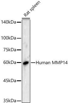
Western blot analysis of lysates from Rat spleen using MMP14/MT1-MMP Rabbit pAb (CAB2549) at 1:500 dilution. Secondary antibody: HRP-conjugated Goat anti-Rabbit IgG (H+L) (CABS014) at 1:10000 dilution. Lysates/proteins: 25 μg per lane. Blocking buffer: 3% nonfat dry milk in TBST. Detection: ECL Basic Kit (AbGn00020). Exposure time: 5s.
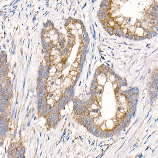
Immunohistochemistry analysis of paraffin-embedded Human colon carcinoma using MMP14/MT1-MMP Rabbit pAb (CAB2549) at dilution of 1:200 (40x lens). High pressure antigen retrieval performed with 0.01M Citrate buffer (pH 6.0) prior to IHC staining.
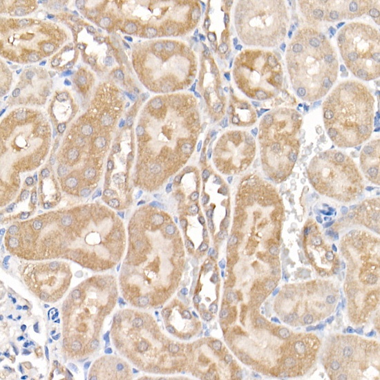
Immunohistochemistry analysis of paraffin-embedded Rat kidney using MMP14/MT1-MMP Rabbit pAb (CAB2549) at dilution of 1:200 (40x lens). High pressure antigen retrieval performed with 0.01M Citrate buffer (pH 6.0) prior to IHC staining.
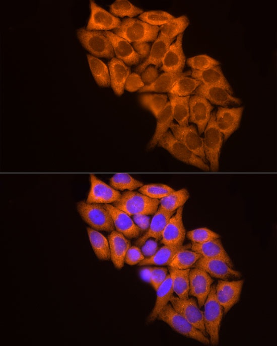
Immunofluorescence analysis of HeLa cells using MMP14/MT1-MMP Rabbit pAb (CAB2549) at dilution of 1:100 (40x lens). Secondary antibody: Cy3-conjugated Goat anti-Rabbit IgG (H+L) (CABS007) at 1:500 dilution. Blue: DAPI for nuclear staining.
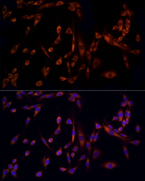
Immunofluorescence analysis of PC-12 cells using MMP14/MT1-MMP Rabbit pAb (CAB2549) at dilution of 1:100 (40x lens). Secondary antibody: Cy3-conjugated Goat anti-Rabbit IgG (H+L) (CABS007) at 1:500 dilution. Blue: DAPI for nuclear staining.


 Polyclonal Antibody (CAB24436)")

