The MTCO2 Antibody (CAB17965) is a high-quality antibody developed for reliable detection and analysis of target proteins. This rabbit polyclonal antibody is highly specific to human samples and has been validated for use in Western blot applications.MT-CO2 is essential for the proper functioning of mitochondria and plays a crucial role in cellular respiration. Dysregulation of MT-CO2 has been implicated in various diseases, including metabolic disorders, neurodegenerative conditions, and cancer.
This antibody is validated for use in WB, IHC-P, IF/ICC, ELISA applications and has demonstrated reactivity against Human, Mouse, Rat samples.
Product Name:
MTCO2 Antibody
SKU:
CAB17965
Size:
20μL, 100μL
Reactivity:
Human, Mouse, Rat
Conjugate:
Unconjugated
Immunogen:
Synthetic peptide. This information is considered to be commercially sensitive.
Sequence:
YALF LTLT TKLT NTNI SDAQ EMET VWTI LPAI ILVL IALP SLRI LYMT DEVN DPSL TIKS I
Tested Applications:
WBIHC-PIF/ICCELISA
Recommended Dilution:
WB
1:1000 - 1:5000
IHC-P
1:50 - 1:200
IF/ICC
1:50 - 1:200
ELISA
Recommended starting concentration is 1 μg/mL. Please optimize the concentration based on your specific assay requirements.
Contributes to cytochrome-c oxidase activity. Predicted to be involved in mitochondrial electron transport, cytochrome c to oxygen and positive regulation of vasoconstriction. Located in mitochondrial inner membrane. Part of respiratory chain complex IV. Biomarker of Huntington's disease and stomach cancer.
Purification Method
Affinity purification
Gene ID
4513
RRID
AB_2861767
Buffer Information
Store at -20℃. Avoid freeze / thaw cycles. Buffer: PBS containing 50% glycerol, preserved with proclin300 or sodium azide, pH 7.3.
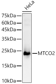
Western blot analysis of lysates from HeLa cells, using MTCO2 Rabbit pAb (CAB17965) at 1:2000 dilution. Secondary antibody: HRP-conjugated Goat anti-Rabbit IgG (H+L) (CABS014) at 1:10000 dilution. Lysates/proteins: 25μg per lane. Blocking buffer: 3% nonfat dry milk in TBST. Detection: ECL Basic Kit (AbGn00020). Exposure time: 10s.
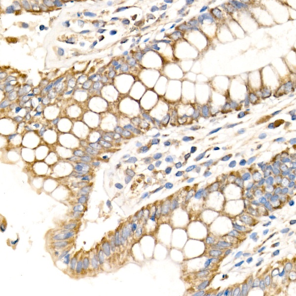
Immunohistochemistry analysis of paraffin-embedded Human colon using MTCO2 Rabbit pAb (CAB17965) at dilution of 1:100 (40x lens). High pressure antigen retrieval performed with 0.01M Citrate buffer (pH 6.0) prior to IHC staining.
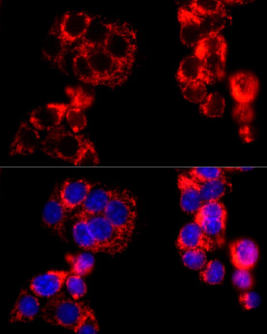
Immunofluorescence analysis of HepG2 cells using MTCO2 Rabbit pAb (CAB17965) at dilution of 1:100 (40x lens). Secondary antibody: Cy3-conjugated Goat anti-Rabbit IgG (H+L) (CABS007) at 1:500 dilution. Blue: DAPI for nuclear staining.