The MMP14/MT1-MMP Monoclonal Antibody (CAB0067) is a high-quality antibody developed for reliable detection and analysis of target proteins. Proteins of the matrix metalloproteinase (MMP) family are involved in the breakdown of extracellular matrix in normal physiological processes, such as embryonic development, reproduction, and tissue remodeling, as well as in disease processes, such as arthritis and metastasis. Most MMP's are secreted as inactive proproteins which are activated when cleaved by extracellular proteinases. However, the protein encoded by this gene is a member of the membrane-type MMP (MT-MMP) subfamily; each member of this subfamily contains a potential transmembrane domain suggesting that these proteins are expressed at the cell surface rather than secreted. This protein activates MMP2 protein, and this activity may be involved in tumor invasion. RRID AB_2861448 Gene ID 4323 Swiss Prot Synonym MMP-14; MMP-X1; MT-MMP; MT1MMP; MTMMP1; WNCHRS; MT1-MMP; MT-MMP 1; MMP14/MT1-MMP
This antibody is validated for use in WB, IHC-P, FC, ELISA applications and has demonstrated reactivity against Human, Mouse, Rat samples.
Product Name:
MMP14/MT1-MMP Monoclonal Antibody
SKU:
CAB0067
Size:
100μL, 20μL
Reactivity:
Human, Mouse, Rat
Clone Number:
ARC0211
Conjugate:
Unconjugated
Immunogen:
Synthetic peptide. This information is considered to be commercially sensitive.
Tested Applications:
WBIHC-PFCELISA
Recommended Dilution:
WB
1:500 - 1:2000
IHC-P
1:50 - 1:200
FC
1:100 - 1:500
ELISA
Recommended starting concentration is 1 μg/mL. Please optimize the concentration based on your specific assay requirements.
HeLa, HepG2, Mouse lung, Mouse kidney, Mouse heart, Rat heart
Cellular Localization:
Cytoplasm, Melanosome, Membrane, Single-Pass Type I Membrane Protein.
Calculated MW:
66kDa
Observed MW:
52kDa/60kDa
Proteins of the matrix metalloproteinase (MMP) family are involved in the breakdown of extracellular matrix in normal physiological processes, such as embryonic development, reproduction, and tissue remodeling, as well as in disease processes, such as arthritis and metastasis. Most MMP's are secreted as inactive proproteins which are activated when cleaved by extracellular proteinases. However, the protein encoded by this gene is a member of the membrane-type MMP (MT-MMP) subfamily; each member of this subfamily contains a potential transmembrane domain suggesting that these proteins are expressed at the cell surface rather than secreted. This protein activates MMP2 protein, and this activity may be involved in tumor invasion. RRID AB_2861448 Gene ID 4323 Swiss Prot Synonym MMP-14; MMP-X1; MT-MMP; MT1MMP; MTMMP1; WNCHRS; MT1-MMP; MT-MMP 1; MMP14/MT1-MMP
Purification Method:
Affinity purification
Gene ID:
4323
RRID:
AB_2861448
Buffer Information:
Store at -20℃. Avoid freeze / thaw cycles. Buffer: PBS containing 50% glycerol and 0.05% BSA, preserved with proclin300 or sodium azide, pH 7.3.
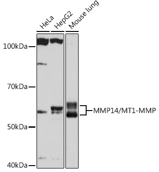
Western blot analysis of various lysates using MMP14/MMP14/MT1-MMP Rabbit mAb (CAB0067) at 1:1000 dilution. Secondary antibody: HRP-conjugated Goat anti-Rabbit IgG (H+L) (AS014) at 1:10000 dilution. Lysates/proteins: 25μg per lane. Blocking buffer: 3% nonfat dry milk in TBST. Detection: ECL Basic Kit (AbGn00020). Exposure time: 90s.
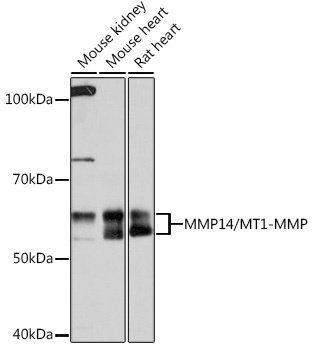
Western blot analysis of various lysates using MMP14/MMP14/MT1-MMP Rabbit mAb (CAB0067) at 1:1000 dilution. Secondary antibody: HRP-conjugated Goat anti-Rabbit IgG (H+L) (AS014) at 1:10000 dilution. Lysates/proteins: 25μg per lane. Blocking buffer: 3% nonfat dry milk in TBST. Detection: ECL Basic Kit (AbGn00020). Exposure time: 10s.
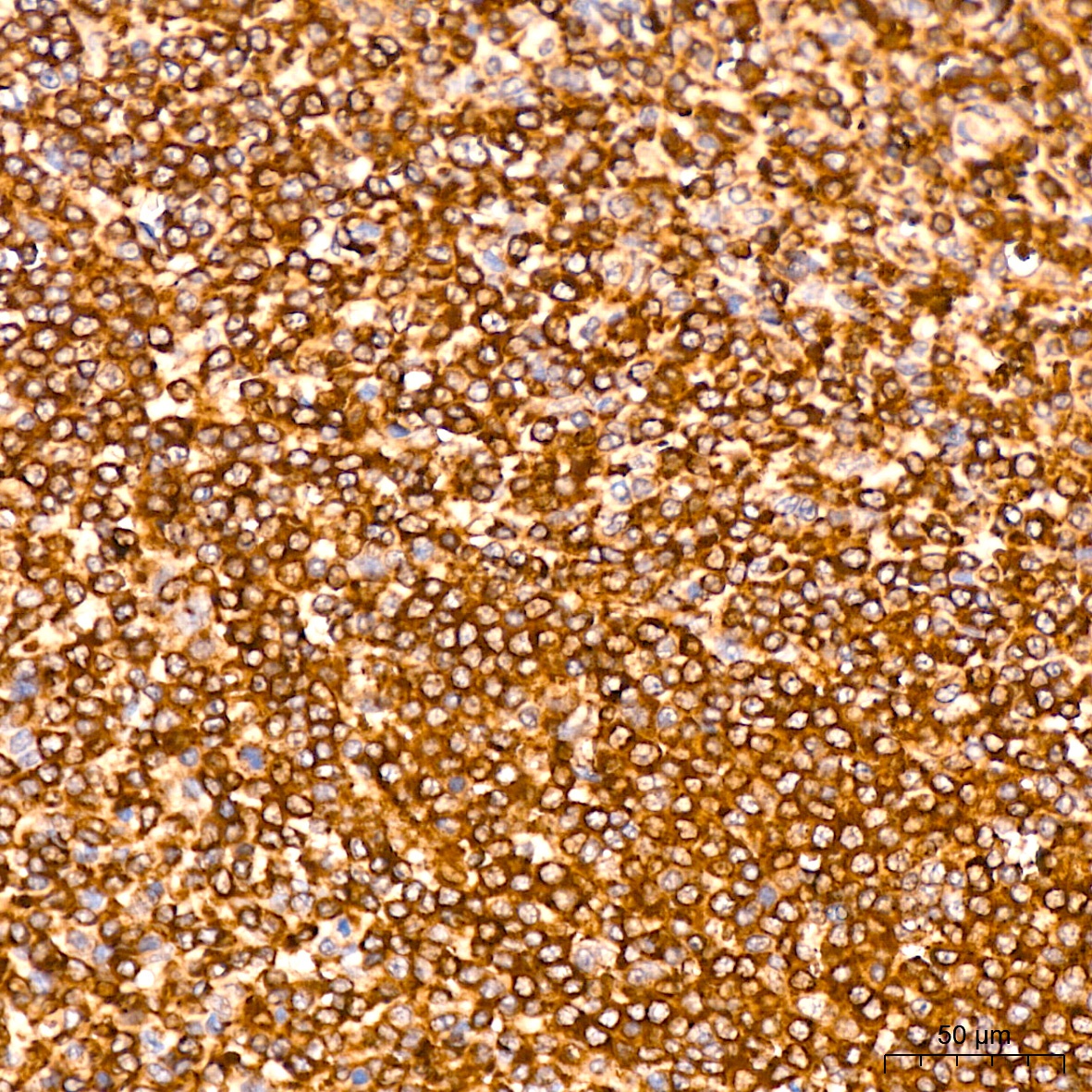
Immunohistochemistry analysis of paraffin-embedded Human spleen tissue using MMP14/MT1-MMP Rabbit mAb (CAB0067) at a dilution of 1:200 (40x lens). High pressure antigen retrieval performed with 0.01M Citrate buffer (pH 6.0) prior to IHC staining.
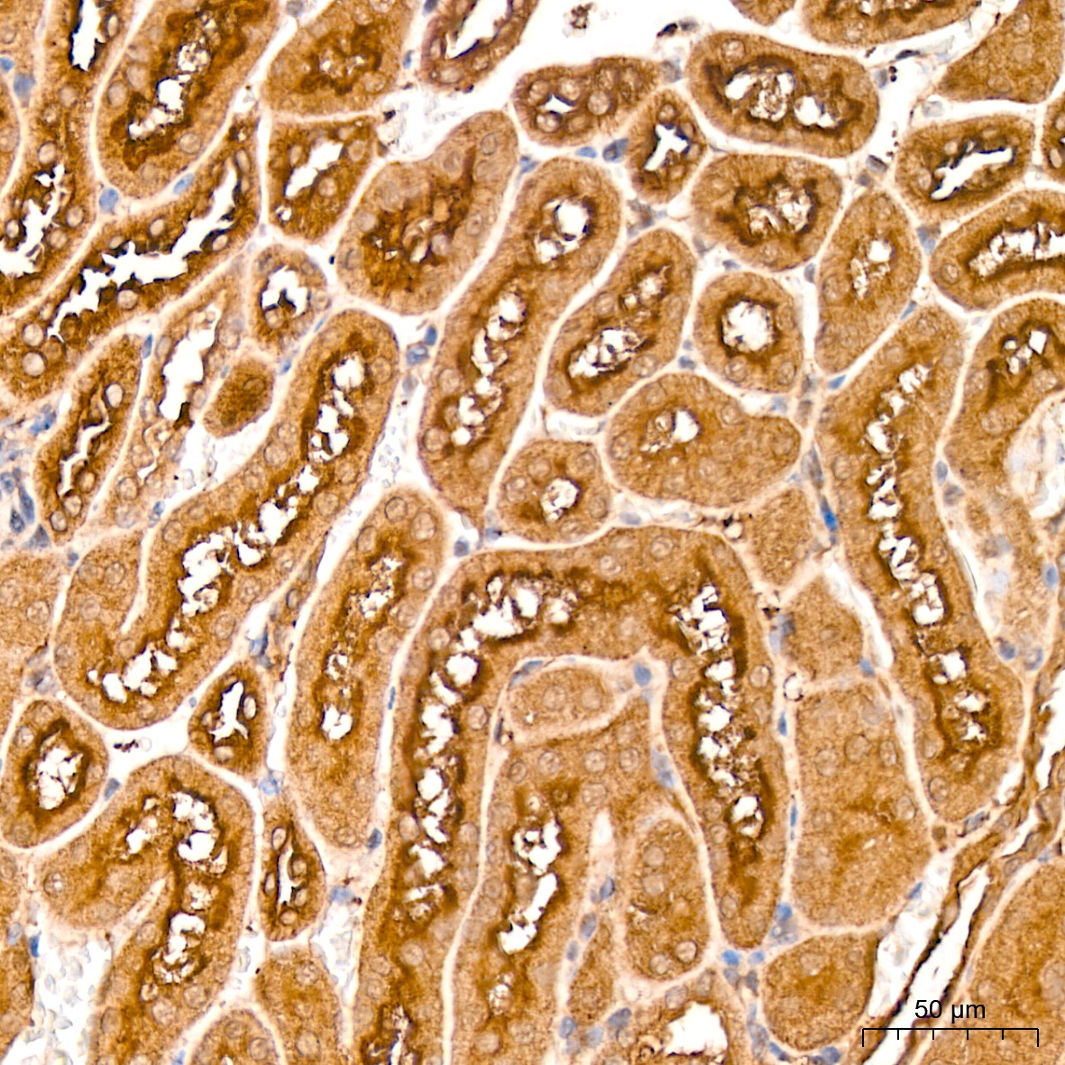
Immunohistochemistry analysis of paraffin-embedded Mouse kidney tissue using MMP14/MT1-MMP Rabbit mAb (CAB0067) at a dilution of 1:200 (40x lens). High pressure antigen retrieval performed with 0.01M Citrate buffer (pH 6.0) prior to IHC staining.
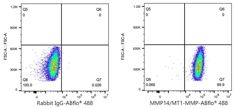
Flow cytometry: 1X10^6 MCF7 cells (negative control,left) and HT-1080 (right) cells were intracellularly-stained with MMP14/MT1-MMP Rabbit mAb (CAB0067,2 μg/mL,orange line) or ABflo® 488 Rabbit IgG isotype control (AC042,2 μg/mL,blue line), followed by Alexa Fluor® 488 conjugated goat anti-rabbit pAb staining. Non-fluorescently stained cells were used as blank control (red line).
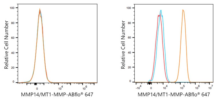
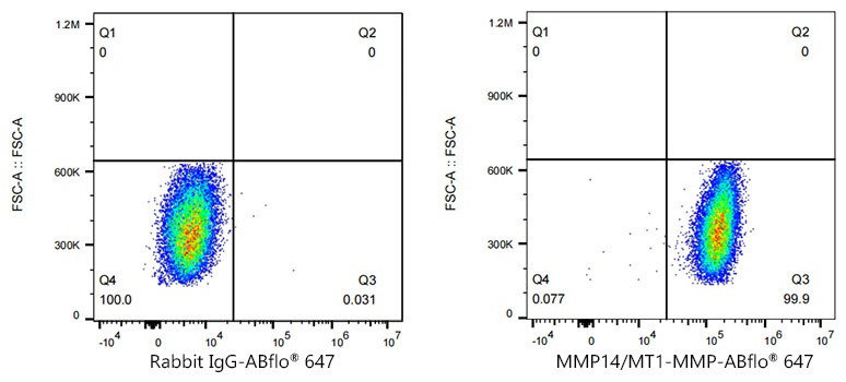
Flow cytometry: 1X10^6 MCF7 cells (negative control,left) and HT-1080 (right) cells were intracellularly-stained with MMP14/MT1-MMP Rabbit mAb (CAB0067,2 μg/mL,orange line) or ABflo® 647 Rabbit IgG isotype control (AC042,2 μg/mL,blue line), followed by Alexa Fluor® 647 conjugated goat anti-rabbit pAb staining. Non-fluorescently stained cells were used as blank control (red line).
Flow cytometry: 1X10^6 MCF7 cells (negative control,left) and HT-1080 (right) cells were intracellularly-stained with MMP14/MT1-MMP Rabbit mAb (CAB0067,2 μg/mL,orange line) or ABflo® 647 Rabbit IgG isotype control (AC042,2 μg/mL,blue line), followed by Alexa Fluor® 647 conjugated goat anti-rabbit pAb staining. Non-fluorescently stained cells were used as blank control (red line).